Spontaneous rupture of the common hepatic duct associated with acute pancreatitis: a case report
- PMID: 28633652
- PMCID: PMC5479041
- DOI: 10.1186/s13256-017-1283-6
Spontaneous rupture of the common hepatic duct associated with acute pancreatitis: a case report
Abstract
Background: Rupture of the common bile duct is a life-threatening condition, usually observed after a trauma or in association with choledocholithiasis or an obstructive tumor of the bile duct. However, a spontaneous rupture of the common bile duct is a rare entity.
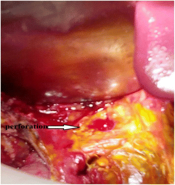
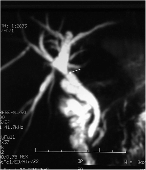
Case presentation: We report a new observation of a spontaneous rupture of the common bile duct, associated with biliary peritonitis and pancreatitis, in a 15-year-old North African girl. Etiological aspects, specificities of clinical presentation, means of diagnosis, as well as surgical and perioperative management are discussed.
Conclusions: The diagnosis of spontaneous rupture of the common bile duct is a challenge for both radiologist and surgeon. Beyond the difficulty of diagnosis, which requires radiological exploration, management of the subsequent biliary peritonitis involves urgent surgery, life-supporting measures, and close monitoring.
Keywords: Cholangiography; Common bile duct; Computed tomography; Pancreatitis; Peritonitis; Spontaneous rupture.
Figures


References
-
- Dijkstra CH. Graluistorting in de buikholtebijeenzuijeling. MaandschrKindergeneesked. 1932;1:409–14.
-
- Roux G, Baumel M, Vidal J, Kornich G. Péritonites biliaires par perforation spontanée. Ann Chir. 1960;14:919–22. - PubMed
Publication types
MeSH terms
Substances
LinkOut - more resources
Full Text Sources
Other Literature Sources
Medical

