. 2017 Jul 17;474(15):2563-2572.
doi: 10.1042/BCJ20170349.

Ascorbate protects the diheme enzyme, MauG, against self-inflicted oxidative damage by an unusual antioxidant mechanism

Affiliations

Ascorbate protects the diheme enzyme, MauG, against self-inflicted oxidative damage by an unusual antioxidant mechanism

Zhongxin Ma et al. Biochem J. .

Abstract

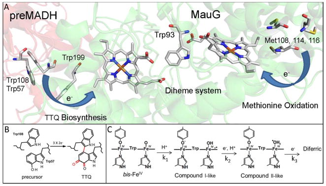
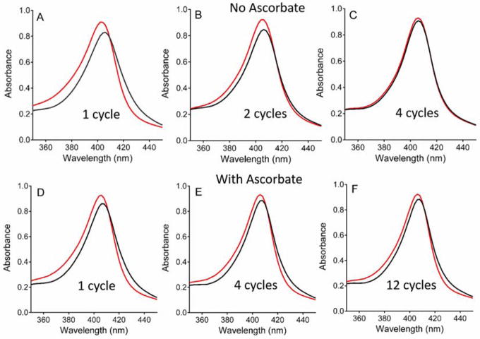
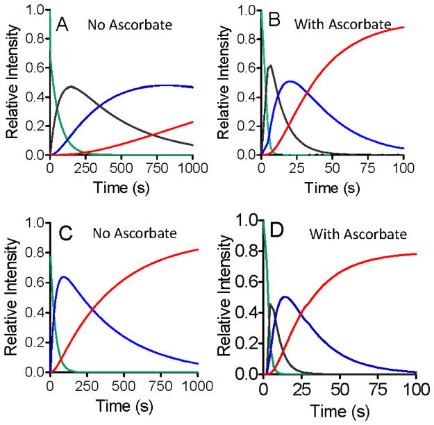
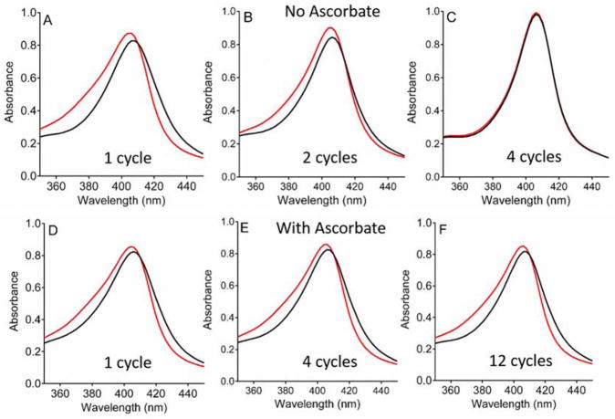
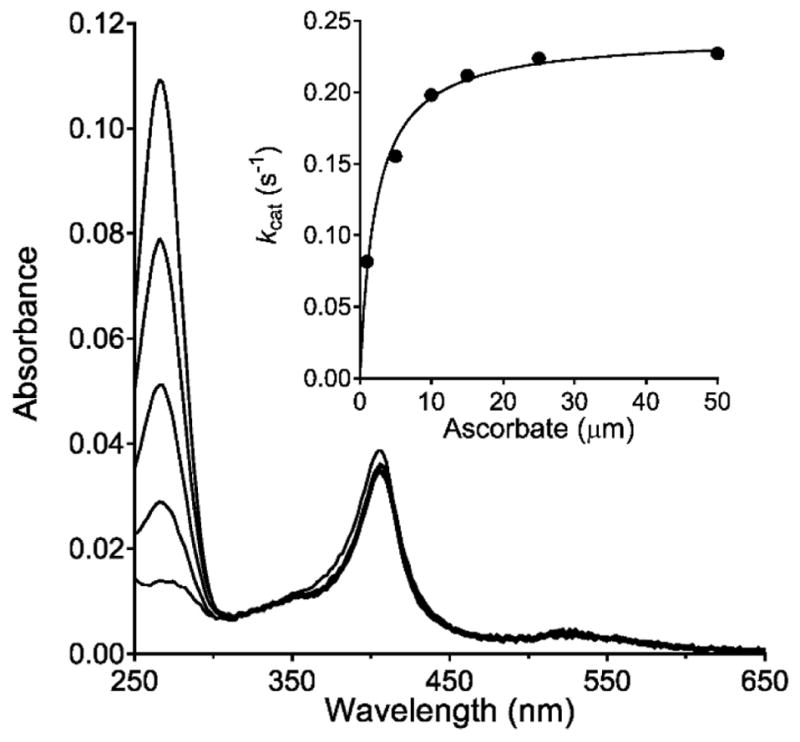
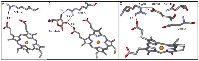
Ascorbate protects MauG from self-inactivation that occurs during the autoreduction of the reactive bis-FeIV state of its diheme cofactor. The mechanism of protection does not involve direct reaction with reactive oxygen species in solution. Instead, it binds to MauG and mitigates oxidative damage that occurs via internal transfer of electrons from amino acid residues within the protein to the high-valent hemes. The presence of ascorbate does not inhibit the natural catalytic reaction of MauG, which catalyzes oxidative post-translational modifications of a substrate protein that binds to the surface of MauG and is oxidized by the high-valent hemes via long-range electron transfer. Ascorbate was also shown to prolong the activity of a P107V MauG variant that is more prone to inactivation. A previously unknown ascorbate peroxidase activity of MauG was characterized with a kcat of 0.24 s-1 and a Km of 2.2 µM for ascorbate. A putative binding site for ascorbate was inferred from inspection of the crystal structure of MauG and comparison with the structure of soybean ascorbate peroxidase with bound ascorbate. The ascorbate bound to MauG was shown to accelerate the rates of both electron transfers to the hemes and proton transfers to hemes which occur during the multistep autoreduction to the diferric state which is accompanied by oxidative damage. A structural basis for these effects is inferred from the putative ascorbate-binding site. This could be a previously unrecognized mechanism by which ascorbate mitigates oxidative damage to heme-dependent enzymes and redox proteins in nature.

Keywords: antioxidants; cytochrome; electron transfer; oxidative stress; peroxidases; proton transfer.


Conflict of interest statement

Declarations of interest

The authors do not have any conflicting financial interests to declare.

Figures

Figure 1
Figure 1
Alternative routes for TTQ biosynthesis and oxidative damage in MauG. A. Relevant portion of the structure of the preMADH-MauG complex (PDB entry 3L4M). The hemes of MauG and amino acid residues referred to in the text are displayed in stick. The background of MauG is green and that of preMADH is pink. B. The posttranslational modification that are catalyzed by MauG to achieve TTQ biosynthesis. C. The intermediates in the autoreduction of the bis-FeIV state to the diferric state. The rate constants associated with each reaction step are indicated.
Figure 2
Figure 2
Changes in the Soret region of the absorption spectrum of WT MauG after redox cycling with H2O2 in the absence of preMADH. The spectra are of MauG before (red) and immediately after addition of stoichiometric H2O2 (black) in the absence of preMADH. Spectral changes in the absence of ascorbate were recorded after one (A), two (B) and four (C) cycles. Spectral changes in the presence of ascorbate were recorded after one (D), four (E) and twelve (F) cycles.
Figure 3
Figure 3
Kinetic plots that depict the global fits of the overall changes in the absorbance spectrum with time that are associated with the conversion of the bis-FeIV state to the diferric state. Experiments were performed with WT MauG in the absence (A) and presence (B) of ascorbate; and with P107V MauG in the absence (C) and presence (D) of ascorbate. These traces correspond to the disappearance of the starting spectrum of the bis-FeIV state (green), the formation and decay of the spectrum of the Compound I-like state when observable (black), the formation and decay of the spectrum of the Compound II-like state (blue) and the formation of the spectrum of the diferric state (red). Spectra were recorded every 2 s.
Figure 4
Figure 4
Changes in the Soret region of the absorption spectrum of P107V MauG after redox cycling with H2O2 in the absence of preMADH. The spectra are of P107V MauG before (red) and immediately after addition of stoichiometric H2O2 (black) in the absence of preMADH. Spectral changes in the absence of ascorbate were recorded after one (A), two (B) and four (C) cycles. Spectral changes in the presence of ascorbate were recorded after one (D), four (E) and twelve (F) cycles.
Figure 5
Figure 5
Steady-state analysis of the ascorbate oxidase activity of MauG. The loss of absorbance of ascorbate (centered at 266 nm) in the presence of MauG and H2O2 was used to monitor ascorbate peroxidase activity. The reaction mixture contained 0.1 μM of MauG and 10 μm ascorbate and the reaction was initiated by addition of 100 μm H2O2. Spectra were recorded at 100 s intervals. The peak centered near 400 nm is from the hemes of MauG. The dependence of the initial rate of reaction on ascorbate concentration with varied concentrations of ascorbate is shown in the inset. The curve is the fit of the data by Eq 1.
Figure 6
Figure 6
Crystal structures of relevant portions of APX and MauG. A. The structure of soybean APX (PDB entry 1OAG). B. The structure of soybean APX in complex with ascorbate (PDB entry 1OAF). C. The structure of MauG (PDB entry 3L4M). Distances in angstroms are indicated next to the dashed lines. The portions of the structures are display as stick with carbon grey, nitrogen blue, oxygen red, and the heme iron as an orange sphere.

Similar articles

Cited by

References

    1. Wang Y, Graichen ME, Liu A, Pearson AR, Wilmot CW, Davidson VL. MauG, a novel diheme protein required for tryptophan tryptophylquinone biogenesis. Biochemistry. 2003;42:7318–7325. - PubMed
    1. Davidson VL, Wilmot CM. Posttranslational biosynthesis of the protein-derived cofactor tryptophan tryptophylquinone. Annu Rev Biochem. 2013;82:531–550. - PMC - PubMed
    1. Pearson AR, de la Mora-Rey T, Graichen ME, Wang Y, Jones LH, Marimanikkupam S, Aggar SA, Grimsrud PA, Davidson VL, Wilmot CW. Further insights into quinone cofactor biogenesis: Probing the role of MauG in methylamine dehydrogenase TTQ formation. Biochemistry. 2004;43:5494–5502. - PubMed
    1. Davidson VL. Pyrroloquinoline quinone (PQQ) from methanol dehydrogenase and tryptophan tryptophylquinone (TTQ) from methylamine dehydrogenase. Adv Protein Chem. 2001;58:95–140. - PubMed
    1. Li X, Fu R, Lee S, Krebs C, Davidson VL, Liu A. A catalytic di-heme bis-Fe(IV) intermediate, alternative to an Fe(IV)=O porphyrin radical. Proc Natl Acad Sci USA. 2008;105:8597–8600. - PMC - PubMed

MeSH terms