Comparative Study
. 2017 Aug 11;292(32):13449-13458.
doi: 10.1074/jbc.M117.787267. Epub 2017 Jun 20.

Structural analyses of human thymidylate synthase reveal a site that may control conformational switching between active and inactive states

Affiliations
Comparative Study

Structural analyses of human thymidylate synthase reveal a site that may control conformational switching between active and inactive states

Dan Chen et al. J Biol Chem. .

Abstract

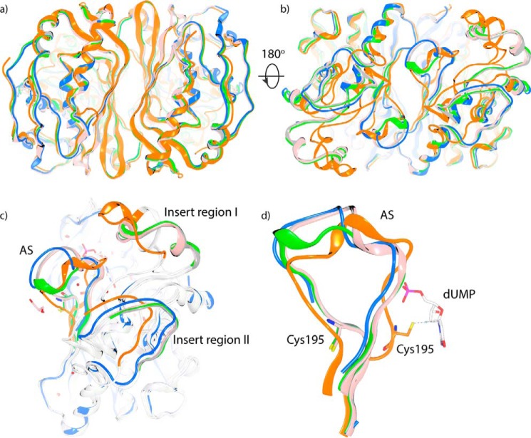
Thymidylate synthase (TS) is the sole enzyme responsible for de novo biosynthesis of thymidylate (TMP) and is essential for cell proliferation and survival. Inhibition of human TS (hTS) has been extensively investigated for cancer chemotherapy, but several aspects of its activity and regulation are still uncertain. In this study, we performed comprehensive structural and biophysical studies of hTS using crystallography and thermal shift assay and provided the first detailed structural information on the conformational changes induced by ligand binding to the hTS active site. We found that upon binding of the antifolate agents raltitrexed and nolatrexed, the two insert regions in hTS, the functions of which are unclear, undergo positional shifts toward the catalytic center. We investigated the inactive conformation of hTS and found that the two insert regions are also involved in the conformational transition between the active and inactive state of hTS. Moreover, we identified a ligand-binding site in the dimer interface, suggesting that the cavity in the dimer interface could serve as an allosteric site of hTS to regulate the conformational switching between the active and inactive states. On the basis of these findings, we propose a regulatory mechanism of hTS activity that involves allosteric regulation of interactions of hTS with its own mRNA depending on cellular demands for TMP.

Keywords: DNA synthesis; antifolate; conformational change; crystallography; drug resistance; nucleotide; thymidylate synthase.


Conflict of interest statement

The authors declare that they have no conflicts of interest with the contents of this article

Figures

Figure 1.
Figure 1.
Characterization of hTS using DSF. a, distinct melting profiles of wild-type and mutant TS proteins. Wild-type TS gives a biphasic melting curve. Mutants M190K and A191K have monophasic melting profiles but give high initial fluorescence. b, dUMP dose-response (DR) data on wild-type TS. Adding dUMP into wild-type TS converts the melting profile to a monophasic curve by stabilizing the first phase. Further titration leads to concentration-dependent stabilization of the monophasic curve. c, dUMP DR data on A191K. d, dUMP DR data on M190K. Addition of dUMP results in reduction of initial fluorescence on both A191K and M190K. The melting profiles of the mutants are gradually transformed to biphasic curves as observed for wild-type TS. A191K is further stabilized at the first phase and exhibits a monophasic profile in the presence of 2 mm dUMP, but M190K fails to achieve that, suggesting M190K has lower affinity for dUMP.
Figure 2.
Figure 2.
Alignment of ligand-free, binary and ternary complex structures. a, overlay of ligand-free structure (blue) and dUMP-bound structure (white). Phosphate ion and dUMP are shown as sticks. b, front view of superposition of protein structures of TS–dUMP (cyan), TS–dUMP–methotrexate (white), TS–dUMP–raltitrexed (yellow), and TS–dUMP–nolatrexed (orange). c, top view of the complex structures. Ligands dUMP and antifolates are shown as stick. d, front view of the folate binding pocket and positional shift of Trp-109.
Figure 3.
Figure 3.
Crystal structures of A191K-sf and A191K-s. a, structural alignment of A191K-sf (gray) and A191K-s (blue). Both are in the inactive conformation with the catalytic cysteine Cys-195 siting outside the active site. b, two phosphate ions bind in the structure of A191K-sf with the 2FoFc density map contoured at 1.0 σ level. c, close up view of the binding site for two conserved phosphate/sulfate ions. P2 is near but different from the phosphate moiety of dUMP (orange). The four arginine residues (Arg-50, Arg-215, Arg-175′, and Arg-176′) that coordinate the phosphate moiety of dUMP undergo positional changes to coordinate the two phosphate ions in the inactive conformation. Upon the conformational changes in the inactive conformation, two distal residues (Arg-185 and Asn-183) in the active conformation move close to the active site and interact with the phosphate ions.
Figure 4.
Figure 4.
Structural comparison of the active and inactive conformations of TS. a, front view of the structural alignment of A191K-sf (gray), A191K-s (pink), M190K (green), TS–dUMP (orange), and the inactive crystal structure of TS (PDB code 1HW3; blue). b, back view of the structural alignment. Significant segmental shifts are observed at this side. c, alignment of the inactive and active conformers with regions undergoing significant conformational changes highlighted. d, structural comparison of the AS loop between the active and inactive states of TS.
Figure 5.
Figure 5.
Binding site identified in the dimer interface. a, crystal structure of F13 binding in the dimer interface of the active hTS conformer. b, electrostatic potential surface for the binding region of F13 in the dimer interface. c, electrostatic potential surface for the region in the inactive conformation corresponding to the binding region of F13. d, superposition of the crystal structure of TS–dUMP (gray) and TS–dUMP–F13 (green) shown in the dimer interface region. Binding of F13 leads to a positional shift of Arg-185 and Arg-185′. The 2FoFc density maps are contoured at the 1.0 σ level around Arg-185 and Arg-185′. e, melting curves of dUMP dose-response on mutant TS R185A. f, dose-response data of F13 on wild-type TS and mutant R185A. Addition of dUMP could drive the protein into the active conformation and also convert the two-phase melting curve into one-phase curve, which facilitates the determination of melting points, therefore the dose-response experiments were carried out in the presence of dUMP. ΔTm values of F13 in the presence of 200 μm dUMP were plotted against the compound concentration from three independent measurements. g, melting curves of the F13 dose-response on A191K. h, melting curves of the F13 dose-response on M190K.

Similar articles

Cited by

References

    1. Hori T., Ayusawa D., Shimizu K., Koyama H., and Seno T. (1984) Chromosome breakage induced by thymidylate stress in thymidylate synthase-negative mutants of mouse FM3A cells. Cancer Res. 44, 703–709 - PubMed
    1. Seno T., Ayusawa D., Shimizu K., Koyama H., Takeishi K., and Hori T. (1985) Thymineless death and genetic events in mammalian cells. Basic Life Sci. 31, 241–263 - PubMed
    1. Wilson P. M., Danenberg P. V., Johnston P. G., Lenz H. J., and Ladner R. D. (2014) Standing the test of time: targeting thymidylate biosynthesis in cancer therapy. Nat. Rev. Clin. Oncol. 11, 282–298 - PubMed
    1. Ackland S. P., Clarke S. J., Beale P., and Peters G. J. (2006) Thymidylate synthase inhibitors. Update Cancer Therap. 1, 403–427 - PubMed
    1. Assaraf Y. (2007) Molecular basis of antifolate resistance. Cancer Metastasis Rev. 26, 153–181 - PubMed

Publication types

MeSH terms