. 2017 Jul 3;114(27):7124-7129.
doi: 10.1073/pnas.1702798114. Epub 2017 Jun 20.

Microbial glycoside hydrolases as antibiofilm agents with cross-kingdom activity

Affiliations

Microbial glycoside hydrolases as antibiofilm agents with cross-kingdom activity

Brendan D Snarr et al. Proc Natl Acad Sci U S A. .

Abstract

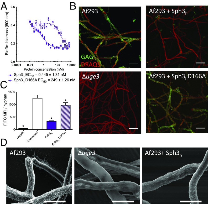
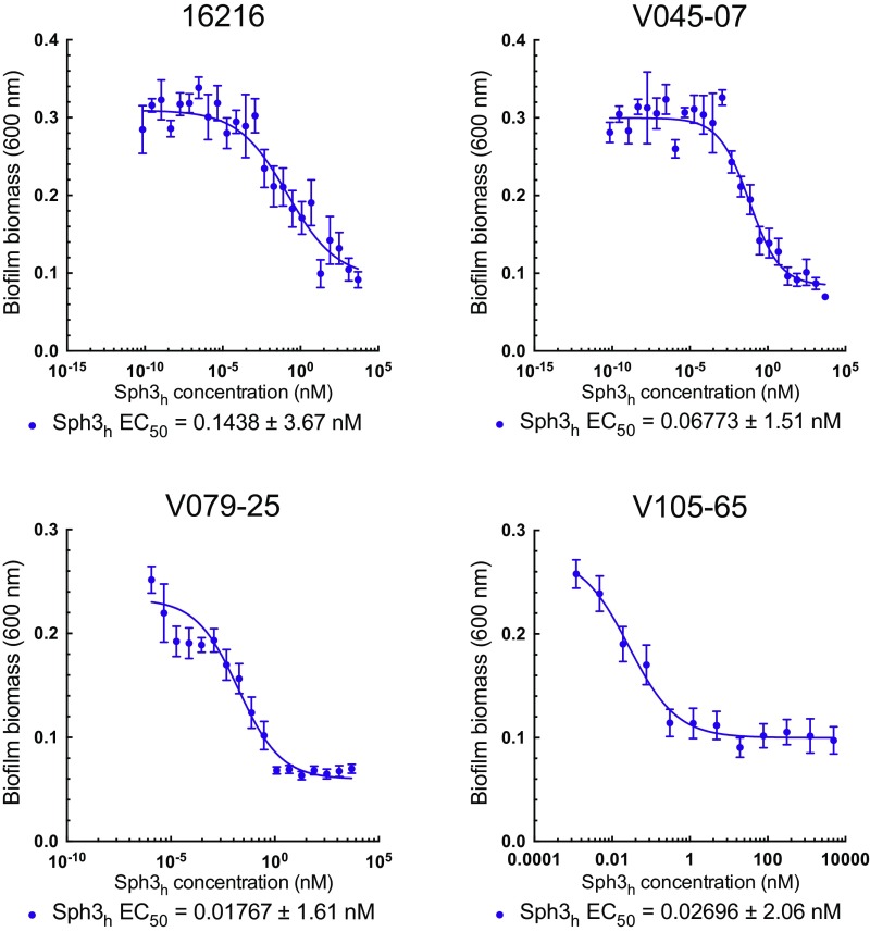
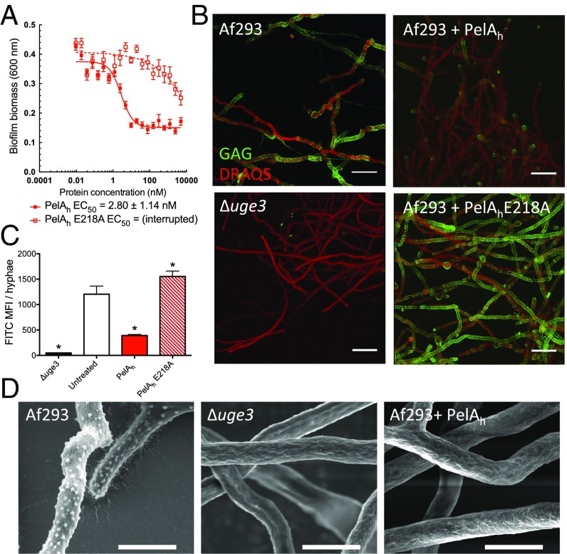
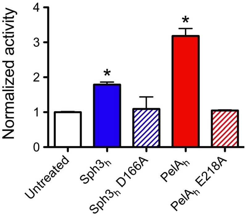
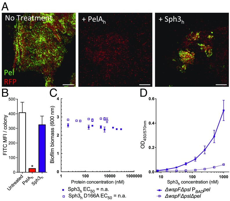
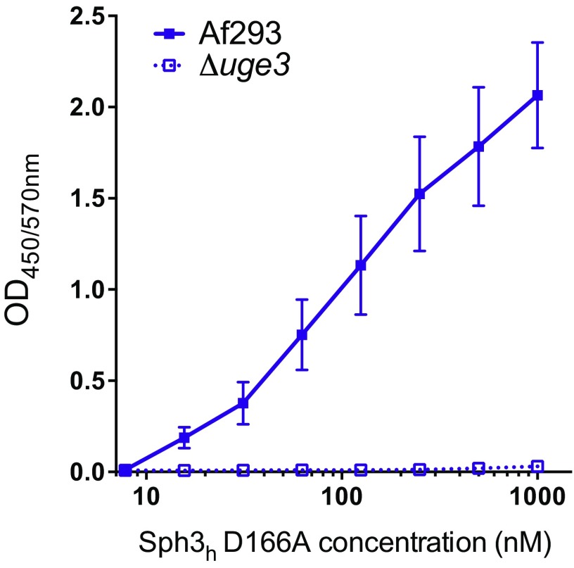
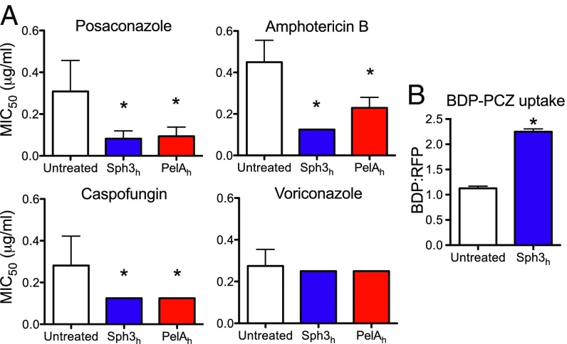
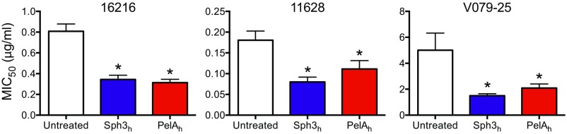
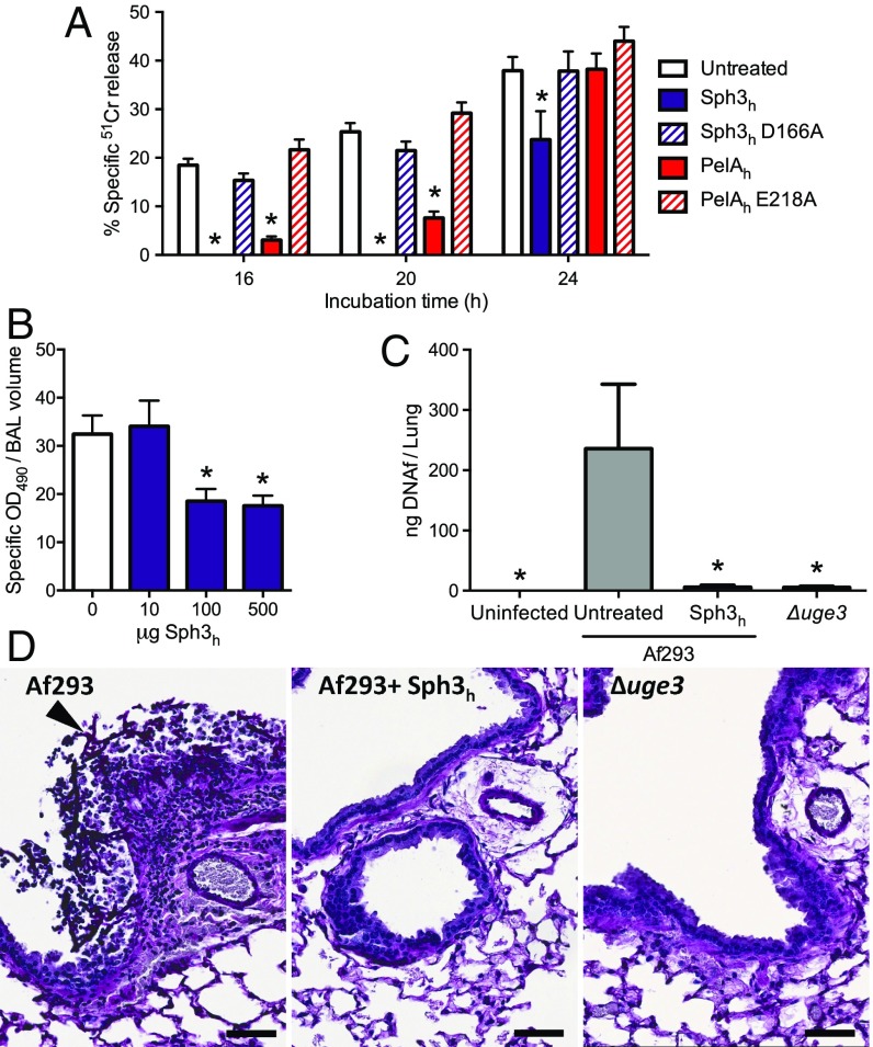
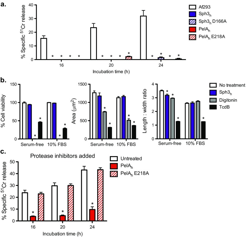
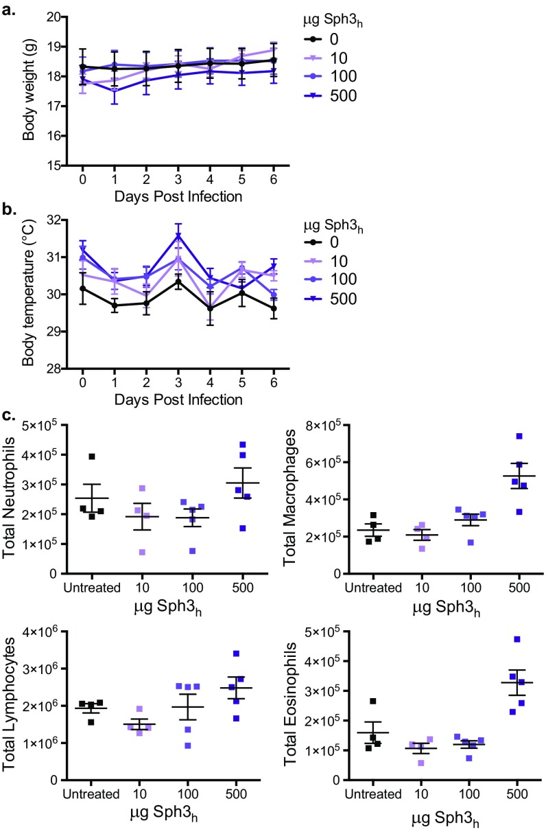
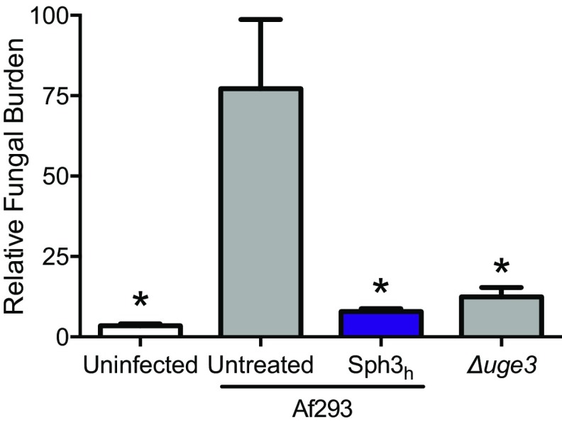
Galactosaminogalactan and Pel are cationic heteropolysaccharides produced by the opportunistic pathogens Aspergillus fumigatus and Pseudomonas aeruginosa, respectively. These exopolysaccharides both contain 1,4-linked N-acetyl-d-galactosamine and play an important role in biofilm formation by these organisms. Proteins containing glycoside hydrolase domains have recently been identified within the biosynthetic pathway of each exopolysaccharide. Recombinant hydrolase domains from these proteins (Sph3h from A. fumigatus and PelAh from P. aeruginosa) were found to degrade their respective polysaccharides in vitro. We therefore hypothesized that these glycoside hydrolases could exhibit antibiofilm activity and, further, given the chemical similarity between galactosaminogalactan and Pel, that they might display cross-species activity. Treatment of A. fumigatus with Sph3h disrupted A. fumigatus biofilms with an EC50 of 0.4 nM. PelAh treatment also disrupted preformed A. fumigatus biofilms with EC50 values similar to those obtained for Sph3h In contrast, Sph3h was unable to disrupt P. aeruginosa Pel-based biofilms, despite being able to bind to the exopolysaccharide. Treatment of A. fumigatus hyphae with either Sph3h or PelAh significantly enhanced the activity of the antifungals posaconazole, amphotericin B, and caspofungin, likely through increasing antifungal penetration of hyphae. Both enzymes were noncytotoxic and protected A549 pulmonary epithelial cells from A. fumigatus-induced cell damage for up to 24 h. Intratracheal administration of Sph3h was well tolerated and reduced pulmonary fungal burden in a neutropenic mouse model of invasive aspergillosis. These findings suggest that glycoside hydrolases can exhibit activity against diverse microorganisms and may be useful as therapeutic agents by degrading biofilms and attenuating virulence.

Keywords: Aspergillus; Pseudomonas; biofilm; exopolysaccharide; therapeutics.


Conflict of interest statement

Conflict of interest statement: A patent has been filed describing the utility of the glycoside hydrolases as antibiofilm therapeutics (CA2951152 A1, WO2015184526 A1). B.D.S., P.B., N.C.B., P.L.H., and D.C.S. are listed as inventors.

Figures

Fig. 1.
Fig. 1.
Treatment with Sph3h disrupts A. fumigatus biofilms and degrades GAG on the surface of hyphae. (A) Crystal violet staining of established A. fumigatus biofilms treated with the indicated concentration of each hydrolase. Each data point represents the mean of n = 20 with error bars indicating SE. EC50 indicates the 50% effective concentration ± SE. (B) Effects of the indicated hydrolases on cell wall-associated GAG. Hyphae of the indicated strains grown in the absence of hydrolase treatment (Left) or following exposure to 0.5 μM of the indicated hydrolases (Right). Cell wall-associated GAG was visualized by using FITC-conjugated lectin staining (green) with DRAQ5 as a counterstain (red). The GAG-deficient Δuge3 mutant was included as a negative control. (C) Quantification of lectin-staining from B. Each data point represents the mean fluorescence intensity of at least seven hyphae with error bars indicating SE. * indicates a significant difference (P < 0.05) relative to the untreated A. fumigatus as determined by one-way ANOVA with Dunnett’s multiple comparison test. (D) Scanning electron micrographs of hyphae of the indicated strains grown in the absence of hydrolases (Left and Center) and following exposure to 0.5 μM Sph3h (Right). (Scale bars: B, 20 μm; D, 5 μm.) MFI, mean fluorescence intensity.
Fig. S1.
Fig. S1.
Effects of Sph3h on biofilms formed by clinical isolates of A. fumigatus. Crystal violet staining of pregrown A. fumigatus biofilms treated with the indicated concentration of Sph3h. Data represents the mean of three independent experiments with error bars indicating SE. EC50 reported is ±SE.
Fig. 2.
Fig. 2.
PelAh disrupts A. fumigatus biofilms and degrades GAG. (A) Effects of PelAh on A. fumigatus biofilms. Crystal violet staining of established A. fumigatus biofilms treated with the indicated concentration of PelAh or PelAh E218A. Each data point represents the mean of n = 20 with error bars indicating SE. EC50 reported ± SE. (B) Effects of the indicated hydrolases on cell wall-associated GAG. Established hyphae of the indicated strains were untreated (Left) or exposed to 0.5 μM of the indicated hydrolases (Right). Cell wall-associated GAG was detected by using FITC-conjugated lectin staining (green), with DRAQ5 as a counterstain (red). (C) Mean fluorescent intensity of lectin staining in B. Each data point represents the mean of at least seven hyphae with error bars indicating SE. The * indicates a significant difference (P < 0.05) relative to the untreated A. fumigatus as determined by one-way ANOVA with Dunnett’s multiple comparison test. (D) Scanning electron micrographs of hyphae of the indicated strains grown in the absence of hydrolase treatment (Left and Center) or following treatment with 0.5 μM PelAh (Right). (Scale bars: B, 20 μm; D, 5 μm.) MFI, mean fluorescence intensity.
Fig. S2.
Fig. S2.
Sph3h and PelAh can hydrolyze purified GAG. Purified GAG was incubated with 12 μM of the indicated hydrolase for 24 h. GAG hydrolysis was measured through quantification of the release of reducing sugars. The * indicates significant difference (P < 0.01) from the untreated samples by unpaired t test. Each bar represents the mean of two independent experiments.
Fig. 3.
Fig. 3.
Sph3h binds Pel but is inactive against established P. aeruginosa biofilm. (A) Established biofilms of RFP-producing P. aeruginosa overexpressing the Pel operon (red) were untreated (Left) or exposed to 0.5 μM of the indicated hydrolases (Center and Right). Biofilm-associated Pel was detected by using FITC-conjugated Wisteria fluoribunda lectin staining (green). (Scale bars: 20 μm.) (B) Mean fluorescent intensity of lectin staining in A. Each data point represents the mean of at least four P. aeruginosa colonies with error bars indicating SE. * indicates a significant difference (P < 0.001) relative to the untreated P. aeruginosa as determined by one-way ANOVA with Dunnett’s multiple comparison test. (C) Effects of Sph3h on Pel-mediated P. aeruginosa biofilms. Crystal violet staining of established biofilms of P. aeruginosa overexpressing the Pel operon incubated with the indicated concentrations of Sph3h or Sph3h D166A. Each data point represents the mean of n = 3 with error bars indicating SE. EC50 reported ± SE. (D) Sph3h binding to Pel polysaccharide. Microtiter plates were coated with culture supernatants of the indicated P. aeruginosa strains and the binding of Sph3h was determined by using an anti-Sph3h antibody. Each data point represents the mean of three independent experiments with error bars indicating SE. MFI, mean fluorescence intensity.
Fig. S3.
Fig. S3.
Sph3h D166A binds to GAG. Microtiter plates were coated with culture supernatants of the indicated A. fumigatus strains, and the binding of the indicated concentrations of Sph3h D166A was determined as above. Data represents the mean of three independent experiments with error bars indicating SE.
Fig. 4.
Fig. 4.
Glycoside hydrolases increase sensitivity of A. fumigatus to antifungal agents. (A) Established biofilms of wild-type A. fumigatus strain Af293 were treated with the indicated concentrations of antifungals with or without 0.5 μM of the indicated hydrolase and the viability of the resulting biofilms was then measured by using the XTT metabolic assay. Susceptibility to antifungals was quantified by determining the antifungal concentration resulting in a 50% reduction in fungal metabolic activity (MIC50) compared with untreated controls. Bars represent the mean of at least n = 4 with error bars indicating SE. (B) Effects of hydrolase therapy on antifungal uptake. A. fumigatus hyphae were treated with 1 μM Sph3h then exposed to 2 μg/mL BDP-PCZ. Uptake of BDP-PCZ was quantified via fluorometry. Each bar represents the mean of three independent experiments with error bars indicating SE. The * indicates a significant difference (P < 0.05) relative to untreated control samples as determined by one-way ANOVA with Dunnett’s multiple comparison test in A, or two-tailed Student’s t test in B. MIC, minimum inhibitory concentration.
Fig. S4.
Fig. S4.
Susceptibility of azole-resistant isolates of A. fumigatus to posaconazole when treated with Sph3h or PelAh. Susceptibility to antifungals was quantified by determining the antifungal concentration resulting in a 50% reduction in fungal metabolic activity (MIC50) compared with untreated controls. The * indicates a significant difference (P < 0.05) from untreated control samples as determined by one-way ANOVA with Dunnett’s multiple comparison test. Each bar represents the mean of at least three independent experiments, with error bars indicating SE. MIC, minimum inhibitory concentration.
Fig. 5.
Fig. 5.
Effects of hydrolases on A. fumigatus-induced airway epithelial cell damage and in vivo pulmonary infection. (A) 51Cr-loaded A549 pulmonary epithelial cells were incubated with conidia of wild-type A. fumigatus in the presence or absence of 0.5 μM concentrations of the indicated hydrolases. Epithelial cell damage was determined by measurement of the amount of 51Cr released into supernatant at the indicated time points. Each bar represents the mean of at least five independent experiments performed in duplicate with error bars indicating SE. (B) Pulmonary injury as measured by lactose dehydrogenase activity of the bronchoalveolar lavage fluid from BALB/c mice treated intratracheally or not with the indicated quantities of Sph3h and killed 7 d after treatment. Data represents the mean of at least n = 5, with error bars representing SE. (C) Fungal burden of neutropenic mice as determined by quantitative PCR following 4 d of infection with the indicated A. fumigatus strain with or without treatment with a single dose of 500 μg of Sph3h. Data represents the mean of at least n = 12, from two independent experiments with error bars indicating SE. The * indicate a significant difference P < 0.01 for A and C, and < 0.05 for B, relative to untreated controls by using a two-way ANOVA for A and a one-way ANOVA for B and C with a Dunnett’s multiple comparison test. (D) Histopathological analysis of lung sections of mice from C stained with periodic acid Schiff reagent. Arrow indicates hyphal lesion. (Scale bars: 20 μm.)
Fig. S5.
Fig. S5.
Sph3h and PelAh are nontoxic to mammalian cell lines in vitro. (A) Chromium-loaded A549 pulmonary epithelial cells were incubated with conidia of wild-type A. fumigatus or 0.5 μM of the indicated hydrolases. Epithelial cell damage was determined by measurement of the amount of chromium released into supernatant at the indicated time points. Each bar represents the mean of at least n = 3, with error bars indicating SE. (B) Potential toxic and morphological effects of the Sph3h on IMR-90 fibroblasts were assayed as previously described. (Left) IMR-90 fibroblast cell viability assay using PrestoBlue reagent. All data were normalized to a no treatment control (100%). IMR-90 cellomics assay to measure the area (Center) and length-to-width ratio (Right) of the cells using CellTracker Orange CMRA. The C. difficile toxin TcdB was used as a positive control in cell morphology assays, and the detergent digitonin was used as a negative control in cell viability assays. Each data point represents the mean from n = 3 from cellomic and PrestoBlue measurements in microtiter plate well, with error bars indicating SE. (C) Chromium-loaded A549 cells were incubated with conidia in the presence or absence of 0.5 μM of the indicated hydrolases and supplemented with 0.1% (vol/vol) protease inhibitor mixture. Each bar represents the mean of four independent experiments performed in triplicate, with error bars indicating SE. The * indicates a significant difference (P < 0.05) compared with cells infected with A. fumigatus alone (A and C) or no treatment (B), as determined by two-way ANOVA with Dunnett’s multiple comparison test.
Fig. S6.
Fig. S6.
Intratracheal administration of Sph3h is well tolerated in BALB/c mice. Body weight (A) and surface body temperature (B) of immunocompetent BALB/c mice administered an intratracheal injection of the indicated amount of Sph3h and monitored daily. Data represents the mean of at least n = 4, with error bars indicating SE. No significance compared with untreated controls, two-way ANOVA with Sidak’s multiple comparisons test. (C) Total pulmonary leukocyte populations of the mice after 7 d posttreatment, as determined by flow cytometry. No significance compared with untreated controls, Kruskal–Wallis test with Dunn’s multiple comparisons test.
Fig. S7.
Fig. S7.
Sph3h coadministration attenuates A. fumigatus invasive infection in BALB/c mice. Fungal burden of neutropenic mice as determined by galactomannan quantification following 4 d of infection with the indicated A. fumigatus strain with or without treatment with a single dose of 500 μg of Sph3h. Data represents the mean of at least n = 12, from two independent experiments with error bars indicating SE. *P < 0.001 compared with untreated mice infected with wild-type A. fumigatus using a one-way ANOVA and Dunnett’s multiple comparison test.

References

    1. Bajwa S, Kulshrestha A. Fungal infections in intensive care unit: Challenges in diagnosis and management. Ann Med Health Sci Res. 2013;3:238–244. - PMC - PubMed
    1. Peleg AY, Hooper DC. Hospital-acquired infections due to gram-negative bacteria. N Engl J Med. 2010;362:1804–1813. - PMC - PubMed
    1. Vincent JL, et al. EPIC International Advisory Committee The prevalence of nosocomial infection in intensive care units in Europe. Results of the European Prevalence of Infection in Intensive Care (EPIC) Study. JAMA. 1995;274:639–644. - PubMed
    1. Maltezou HC. Metallo-β-lactamases in Gram-negative bacteria: Introducing the era of pan-resistance? Int J Antimicrob Agents. 2009;33:405.e1–405.e7. - PubMed
    1. Brown GD, et al. Hidden killers: Human fungal infections. Sci Transl Med. 2012;4:165rv13. - PubMed

Publication types