. 2017:2017:4814987.
doi: 10.1155/2017/4814987. Epub 2017 May 28.

Characterization of the Proinflammatory Profile of Synovial Fluid-Derived Exosomes of Patients with Osteoarthritis

Affiliations

Characterization of the Proinflammatory Profile of Synovial Fluid-Derived Exosomes of Patients with Osteoarthritis

Rossana Domenis et al. Mediators Inflamm. 2017.

Abstract

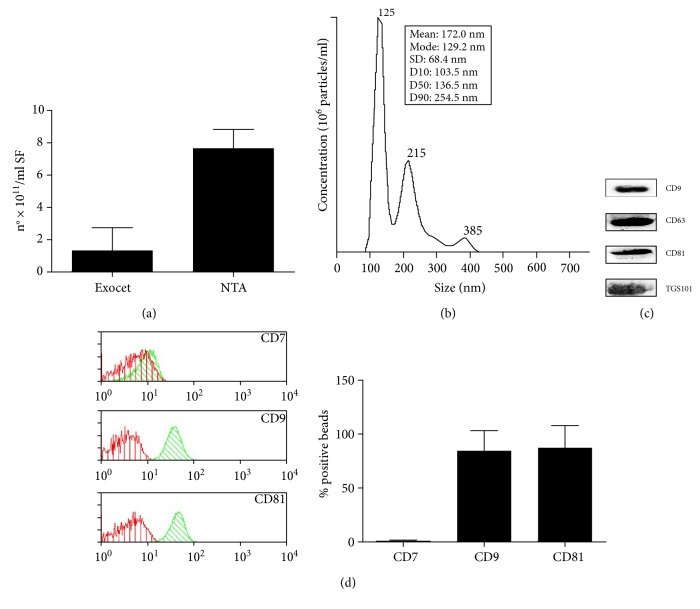
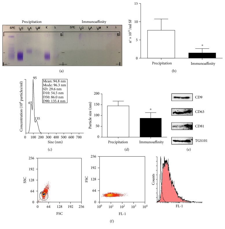
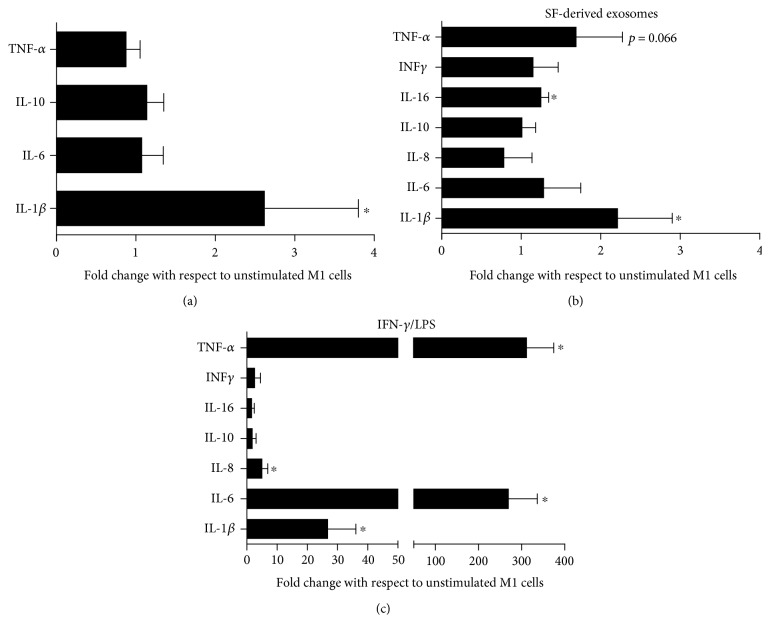
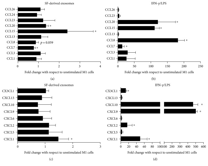
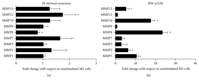
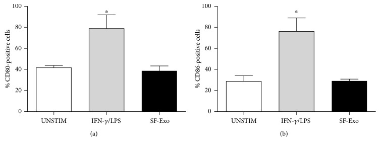
The purpose of this study is to characterize synovial fluid- (SF-) derived exosomes of patients with gonarthrosis comparing two methods of isolation and to investigate their immune regulatory properties. Extracellular vesicles (EVs) have been isolated from inflamed SF by polymer precipitation method and quantified by Exocet kit and by nanoparticle tracking analysis. Vesicles expressed all the specific exosomal markers by immunoblot and FACS. After isolation with Exoquick, a relevant contamination by immune complexes was detected, which required further magnetic bead-based purification to remove. SF-derived exosomes significantly stimulated the release of several inflammatory cytokines and chemokines and metalloproteinases by M1 macrophages but did not influence the expression of CD80 and CD86 costimulatory molecules. In conclusion, we characterized purified exosomes isolated from inflamed SF and demonstrate that purified exosomes are functionally active in their ability to stimulate the release of proinflammatory factors from M1 macrophages. Our data indicate that SF-derived exosomes from gonarthrosis patients play a role in disease progression.


Figures

Figure 1
Figure 1
Characterization of SF-derived EVs isolated by Exoquick polymer precipitation. (a) Particle concentration was quantified measuring the enzymatic activity of the exosomal AChE enzyme by Exocet kit or tracking the particles' Brownian motion by Nanosight NTA. Columns, mean; bars, SD. (b) A representative particle size profiling by NTA is shown. (c) SF-derived EVs were lysed, and 25 μg of protein was separated in 15% SDS-PAGE gel under reducing condition. The gel was western blotted onto nitrocellulose membranes and stained with antibodies recognizing exosomal marker proteins CD9, CD63, CD81, and TSG101. (d) EVs were embedded into 4 μm beads coated with anti-CD63 and then stained with specific monoclonal antibody for CD7, CD9, and CD81 and analysed by flow cytometry. The antibodies (green peak) were compared with their appropriate isotype control (red peak). Cytometry histograms are shown as one representative experiment. The histograms represent the percentages of CD7-, CD9-, and CD81-positive beads. Columns, mean; bars, SD.
Figure 2
Figure 2
Characterization of SF-derived exosomes isolated by immunoaffinity. (a) SF-derived exosomes isolated by Exoquick polymer precipitation or immunoaffinity magnetic beads were assessed for immune complex contamination by immunofixation. The immunofixation gel is shown as one representative experiment. The particle concentration (b) and size (d) were quantified tracking the particles' Brownian motion by Nanosight NTA. Columns, mean; bars, SD, significant difference p < 0.05. (c) A representative particle size profiling by NTA is shown. (e) SF-derived exosomes were lysed, and 2 μg of protein was separated in 15% SDS-PAGE gel under reducing condition. The gel was blotted onto nitrocellulose membranes and stained with antibodies against exosomal markers CD9, CD63, CD81, and TSG101. (f) Exosomes were bound by Exo-Flow beads, stained with Exo-FITC, and analysed by flow cytometry. Histograms of one representative experiment are shown. Exosome-bound beads (red peak) were compared with beads alone (white peak).
Figure 3
Figure 3
SF-derived exosomes stimulate the production of several cytokines by M1 macrophages. (a) M1 macrophages, differentiated from normal donor monocytes, were incubated with SF-derived exosomes for 6 hours and cytokine expression was analysed by RT-PCR. Data were expressed as fold change relative to unstimulated cells. (b) M1 macrophages were treated with SF-derived exosomes (b) or IFN-γ/LPS (c) for 24 hours, and cytokine production was quantified in supernatants by ELISA Bio-plex cytokine assay system. Data were expressed as fold change relative to unstimulated cells. Columns, mean; bars, SD, significant difference from unstimulated cells p < 0.05.
Figure 4
Figure 4
SF-derived exosomes stimulate the production of several chemokines by M1 macrophages. M1 macrophages were treated with SF-derived exosomes (a–c) or IFN-γ/LPS (b–d) for 24 h, and chemokine production in supernatants was determined by the ELISA Bio-plex chemokines assay system. Data are displayed as fold change with respect to unstimulated cells. Columns, mean; bars, SD, significant difference from unstimulated cells p < 0.05.
Figure 5
Figure 5
SF-derived exosomes stimulate the production of several MMPs by M1 macrophages. M1 macrophages were treated with SF-derived exosomes (a) or IFN-γ/LPS (b) for 24 h, and MMP production was determined in supernatants by the ELISA Bio-plex assay system. Data are expressed as fold change in relation to unstimulated cells. Columns, mean; bars, SD, significant difference from unstimulated cells p < 0.05.
Figure 6
Figure 6
Expression of CD80 and CD86 by M1 macrophages following exposure to SF-derived exosomes. M1 macrophages were incubated in the presence of IFN-γ/LPS (grey column) or SF-derived exosomes (black column), and the percentage of CD80- (a) and CD86- (b) positive cells was determined by flow cytometry after 24 hours. Columns, mean; bars, SD. significant difference from unstimulated cells p < 0.05.

References

    1. Malemud C. J. Biologic basis of osteoarthritis: state of the evidence. Current Opinion in Rheumatology. 2015;27(3):289–294. doi: 10.1097/BOR.0000000000000162. - DOI - PMC - PubMed
    1. de Lange-Brokaar B. J., Ioan-Facsinay A., van Osch G. J., et al. Synovial inflammation, immune cells and their cytokines in osteoarthritis: a review. Osteoarthritis and Cartilage. 2012;20(12):1484–1499. doi: 10.1016/j.joca.2012.08.027. - DOI - PubMed
    1. Haywood L., McWilliams D. F., Pearson C. I., et al. Inflammation and angiogenesis in osteoarthritis. Arthritis and Rheumatism. 2003;48(8):2173–2177. doi: 10.1002/art.11094. - DOI - PubMed
    1. Kennedy A., Fearon U., Veale D. J., Godson C. Macrophages in synovial inflammation. Frontiers in Immunology. 2011;2:p. 52. doi: 10.3389/fimmu.2011.00052. - DOI - PMC - PubMed
    1. Bondeson J., Blom A. B., Wainwright S., Hughes C., Caterson B., van den Berg W. B. The role of synovial macrophages and macrophage-produced mediators in driving inflammatory and destructive responses in osteoarthritis. Arthritis and Rheumatism. 2010;62(3):647–657. doi: 10.1002/art.27290. - DOI - PubMed