. 2017 Jun 6:8:644.
doi: 10.3389/fimmu.2017.00644. eCollection 2017.

Whole Blood Profiling of Bacillus Calmette-Guérin-Induced Trained Innate Immunity in Infants Identifies Epidermal Growth Factor, IL-6, Platelet-Derived Growth Factor-AB/BB, and Natural Killer Cell Activation

Affiliations

Whole Blood Profiling of Bacillus Calmette-Guérin-Induced Trained Innate Immunity in Infants Identifies Epidermal Growth Factor, IL-6, Platelet-Derived Growth Factor-AB/BB, and Natural Killer Cell Activation

Steven G Smith et al. Front Immunol. .

Abstract

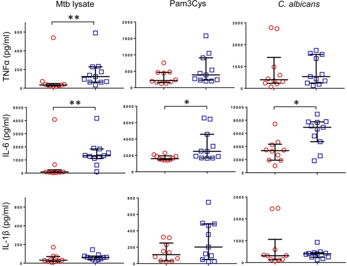
Vaccination of infants with bacillus Calmette-Guérin (BCG) activates both the innate and adaptive arms of the immune response. The antimycobacterial effects of these responses most likely account for the ability of BCG to protect against childhood forms of tuberculosis (TB). There is also evidence for a heterologous protective effect of BCG vaccination against TB-unrelated mortality in low birth weight infants. A possible mechanism of action of this effect, the induction of trained innate immunity, has been demonstrated when cells from BCG-vaccinated adults are restimulated in vitro with non-related microbial stimuli. Our aim was to examine an extensive panel of secreted immune biomarkers to characterize the profile of trained innate immunity in infants. Stimulation of whole blood for 48 h was performed 4 months after BCG vaccination, or in control unvaccinated infants. Stimulants were lipopolysaccharide; Pam3Cys (P3C); heat-killed Candida albicans, Staphylococcus aureus, Escherichia coli, and a lysate of Mycobacterium tuberculosis. Culture supernatants were tested for secreted cytokines and chemokines by 42-plex bead array and monocytes and natural killer (NK) cells assessed for expression of activation markers by flow cytometry. BCG-vaccinated infants displayed increases in 11 cytokines and chemokines in response to different non-specific innate immunity stimuli: epidermal growth factor (EGF); eotaxin; IL-6; IL-7; IL-8; IL-10; IL-12p40; monocyte chemotactic protein-3; macrophage inflammatory protein-1α; soluble CD40 ligand and platelet-derived growth factor (PDGF)-AB/BB. Although each stimulant induced a distinct response profile, three analytes, EGF, IL-6, and PDGF-AB/BB, were commonly higher after stimulation with Pam3Cys, C. albicans, and S. aureus. Conversely, certain cytokines such as interferon gamma-inducible protein-10, IL-2, IL-13, IL-17, GM-CSF, and GRO were suppressed in BCG-vaccinated infants, while no increases in TNFα or IL-1β production were detected. We did not observe a concomitant, BCG-associated change in monocyte surface activation markers in response to non-specific stimuli, but we detected a significant increase in CD69 expression on NK cells in response to Pam3Cys. Pam3Cys-induced NK cell activation correlated with the magnitude of IL-12p40 and IL-10 responses to the same stimulant. This study reveals a novel cytokine/chemokine biomarker signature of BCG-induced trained innate immunity in infants and the involvement of NK cells in these responses.

Keywords: bacillus Calmette–Guérin; chemokines; cytokines; heterologous effects; infants; natural killer; trained immunity; vaccination.


Figures

Figure 1
Figure 1
Cytokine profile of bacillus Calmette–Guérin (BCG)-induced trained immunity in infants does not include TNFα or IL-1β. TNFα, IL-6, and IL-1β concentrations were measured in tissue culture supernatants after diluted whole blood from BCG-vaccinated (□, n = 11) and unvaccinated (○, n = 10) infants was cultured for 48 h with the indicated stimulants. Data shown is background subtracted (unstimulated samples). Wide bars indicate median response; narrow bars indicate 25th and 75th percentiles. Responses in vaccinated and unvaccinated groups were compared using the Mann–Whitney U test: **p < 0.01; *p < 0.05.
Figure 2
Figure 2
Distinct patterns of cytokine and chemokine secretion characterize bacillus Calmette–Guérin (BCG)-induced trained immunity in response to different, non-specific stimuli. The concentrations of 42 cytokines and chemokines were measured in 48 h, diluted whole blood assay supernatants using multiplex bead array. (A) Heat maps represent the vaccination effect [the log2 median fold change in analyte concentration in the BCG-vaccinated infants (n = 11) compared to unvaccinated infants (n = 10)] on the indicated cytokine/chemokine responses to different stimuli: ML, Mtb lysate; LPS, lipopolysaccharide; EC, E. coli; P3C, Pam3Cys; CA, C. albicans; SA, S. aureus. (B) Volcano plots represent the vaccination effect on cytokine/chemokine responses to different indicated stimuli which are plotted on the x-axis against the −log10 p value on the y-axis (calculated by Mann–Whitney U test comparisons of responses in BCG-vaccinated and unvaccinated groups). Data points represent individual cytokines and chemokines. Significant responses of interest are labeled where clarity permits. Horizontal dotted lines represent a p value of 0.05.
Figure 2
Figure 2
Distinct patterns of cytokine and chemokine secretion characterize bacillus Calmette–Guérin (BCG)-induced trained immunity in response to different, non-specific stimuli. The concentrations of 42 cytokines and chemokines were measured in 48 h, diluted whole blood assay supernatants using multiplex bead array. (A) Heat maps represent the vaccination effect [the log2 median fold change in analyte concentration in the BCG-vaccinated infants (n = 11) compared to unvaccinated infants (n = 10)] on the indicated cytokine/chemokine responses to different stimuli: ML, Mtb lysate; LPS, lipopolysaccharide; EC, E. coli; P3C, Pam3Cys; CA, C. albicans; SA, S. aureus. (B) Volcano plots represent the vaccination effect on cytokine/chemokine responses to different indicated stimuli which are plotted on the x-axis against the −log10 p value on the y-axis (calculated by Mann–Whitney U test comparisons of responses in BCG-vaccinated and unvaccinated groups). Data points represent individual cytokines and chemokines. Significant responses of interest are labeled where clarity permits. Horizontal dotted lines represent a p value of 0.05.
Figure 3
Figure 3
Bacillus Calmette–Guérin (BCG) vaccination of infants is associated with enhanced natural killer (NK) cell activation in response to Pam3Cys. Following 48 h, diluted whole blood assay stimulation of samples from BCG-vaccinated (□, n = 10) and unvaccinated (○, n = 8) infants, cell pellets were cryopreserved and later analyzed by flow cytometry. (A) Monocytes were identified based on high CD14 and HLA-DR expression as well as on size and granularity (see Figure S1A in Supplementary Material) and stained for the indicated markers of monocyte activation and differentiation. Data points represent the fold change in MFI for samples incubated with the indicated stimulants compared to unstimulated samples. (B) CD56+ CD3− NK cells were gated (see Figure S1B in Supplementary Material) and the expression of the activation markers CD69 and CD25 analyzed. Data points represent the fold change in percent of activation marker expressing NK cells for samples incubated with the indicated stimulants compared to unstimulated samples. Responses in vaccinated and unvaccinated groups were compared using the Mann–Whitney U test: *p < 0.05; **p < 0.01.
Figure 4
Figure 4
Correlation between Pam3Cys (P3C)-induced natural killer (NK) cell CD69 expression and IL-12p40 and IL-10 secretion. P3C-specific, NK cell CD69 expression in samples from bacillus Calmette–Guérin (BCG)-vaccinated (□, n = 10) and unvaccinated (○, n = 8) infants were plotted against P3C-specific secretion of IL-12p40 and IL-10 in the same samples. The degree of correlation was measured using Spearman’s rank correlation coefficient.

References

    1. Colditz GA, Brewer TF, Berkey CS, Wilson ME, Burdick E, Fineberg HV, et al. Efficacy of BCG vaccine in the prevention of tuberculosis. Meta-analysis of the published literature. JAMA (1994) 271:698–702.10.1001/jama.1994.03510330076038 - DOI - PubMed
    1. Mangtani P, Abubakar I, Ariti C, Beynon R, Pimpin L, Fine PEM, et al. Protection by BCG vaccine against tuberculosis: a systematic review of randomized controlled trials. Clin Infect Dis (2014) 58:470–80.10.1093/cid/cit790 - DOI - PubMed
    1. Wilson ME, Fineberg HV, Colditz GA. Geographic latitude and the efficacy of bacillus Calmette-Guérin vaccine. Clin Infect Dis (1995) 20:982–91.10.1093/clinids/20.4.982 - DOI - PubMed
    1. Colditz GA, Berkey CS, Mosteller F, Brewer TF, Wilson ME, Burdick E, et al. The efficacy of bacillus Calmette-Guérin vaccination of newborns and infants in the prevention of tuberculosis: meta-analyses of the published literature. Pediatrics (1995) 96:29–35. - PubMed
    1. Black GF, Weir RE, Floyd S, Bliss L, Warndorff DK, Crampin AC, et al. BCG-induced increase in interferon-gamma response to mycobacterial antigens and efficacy of BCG vaccination in Malawi and the UK: two randomised controlled studies. Lancet (2002) 359:1393–401.10.1016/S0140-6736(02)08353-8 - DOI - PubMed