. 2017 May 28;9(5):230-244.
doi: 10.4329/wjr.v9.i5.230.

Cystic lesions of peripheral nerves: Are we missing the diagnosis of the intraneural ganglion cyst?

Affiliations

Cystic lesions of peripheral nerves: Are we missing the diagnosis of the intraneural ganglion cyst?

Jyoti Panwar et al. World J Radiol. .

Abstract

Aim: To highlight the salient magnetic resonance imaging (MRI) features of the intraneural ganglion cyst (INGC) of various peripheral nerves for their precise diagnosis and to differentiate them from other intra and extra-neural cystic lesions.

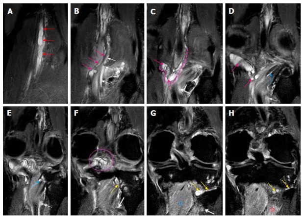
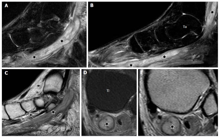
Methods: A retrospective analysis of the magnetic resonance (MR) images of a cohort of 245 patients presenting with nerve palsy involving different peripheral nerves was done. MR images were analyzed for the presence of a nerve lesion, and if found, it was further characterized as solid or cystic. The serial axial, coronal and sagittal MR images of the lesions diagnosed as INGC were studied for their pattern and the anatomical extent along the course of the affected nerve and its branches. Its relation to identifiable anatomical landmarks, intra-articular communication and presence of denervation changes in the muscles supplied by involved nerve was also studied.

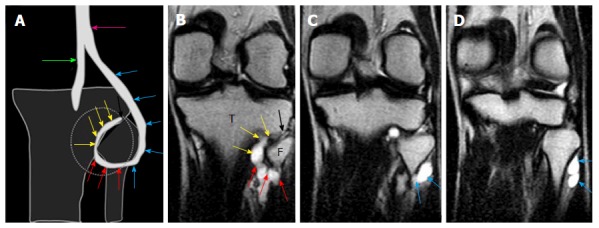
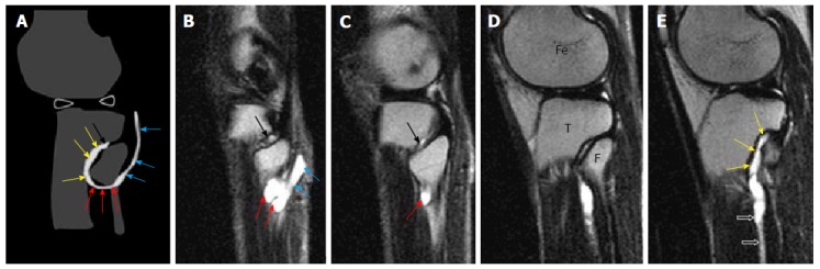
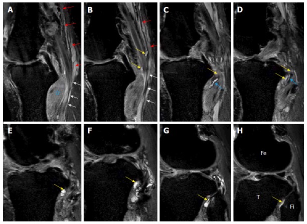
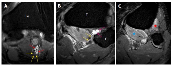
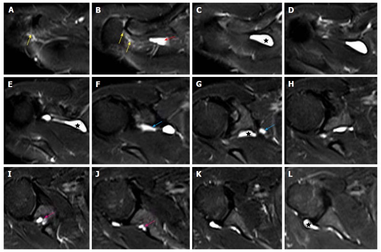
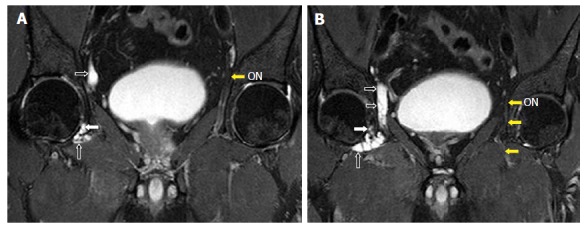
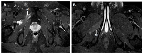
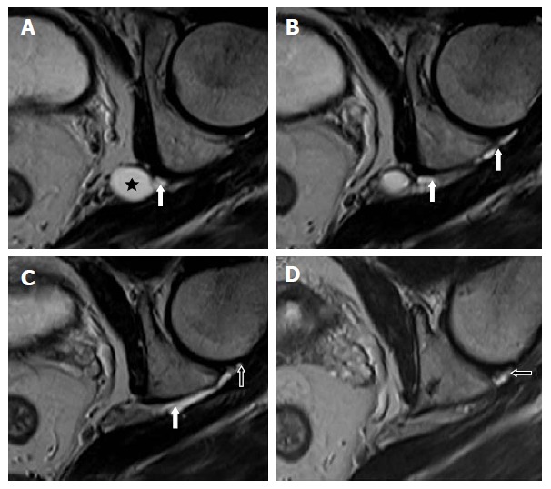
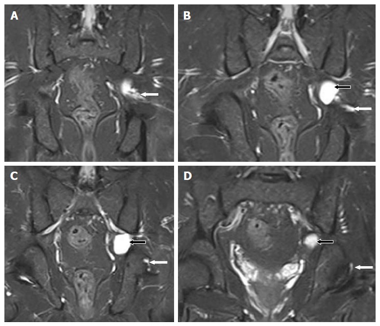
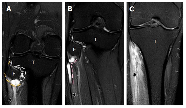
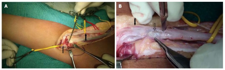
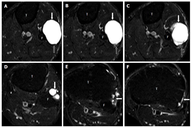
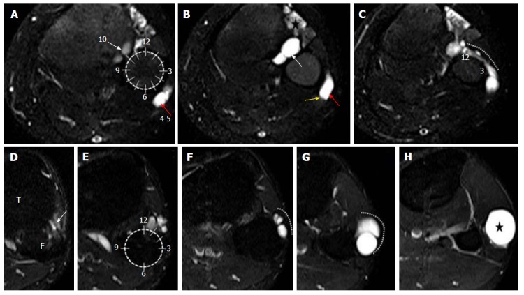
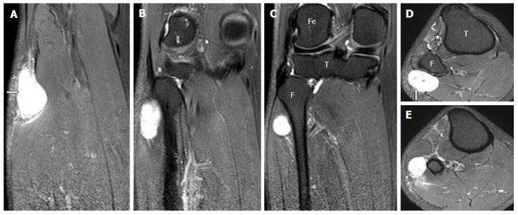
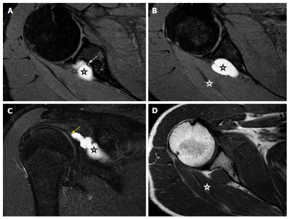
Results: A total of 45 cystic lesions in the intra or extraneural locations of the nerves were identified from the 245 MR scans done for patients presenting with nerve palsy. Out of these 45 cystic lesions, 13 were diagnosed to have INGC of a peripheral nerve on MRI. The other cystic lesions included extraneural ganglion cyst, paralabral cyst impinging upon the suprascapular nerve, cystic schwannoma and nerve abscesses related to Hansen's disease involving various peripheral nerves. Thirteen lesions of INGC were identified in 12 patients. Seven of these affected the common peroneal nerve with one patient having a bilateral involvement. Two lesions each were noted in the tibial and suprascapular nerves, and one each in the obturator and proximal sciatic nerve. An intra-articular connection along the articular branch was demonstrated in 12 out of 13 lesions. Varying stages of denervation atrophy of the supplied muscles of the affected nerves were seen in 7 cases. Out of these 13 lesions in 12 patients, 6 underwent surgery.

Conclusion: INGC is an important cause of reversible mono-neuropathy if diagnosed early and surgically treated. Its classic MRI pattern differentiates it from other lesions of the peripheral nerve and aid in its therapeutic planning. In each case, the joint connection has to be identified preoperatively, and the same should be excised during surgery to prevent further cyst recurrence.

Keywords: Extra-neural; Ganglion cyst; Intra-neural; Magnetic resonance imaging; Peripheral nerves.


Conflict of interest statement

Conflict-of-interest statement: There are no conflicts of interest to report.

Figures

Figure 1
Figure 1
A shows a diagrammatic representation of the intraneural ganglion cyst associated with the proximal tibiofibular joint in the coronal plane; B-D are serial, coronal, T2-weighted, fast spin echo images of the knee show the origin of the lobulated tubular cyst from the proximal tibiofibular joint also called the “tail sign” demonstrated by the black arrows. The further extension along the descending limb (yellow arrows) of the articular branch represents the “vertical limb sign”. The ascending limb of the articular branch (red arrows) demonstrates the “transverse limb sign” which continues to the CPN (blue arrows). Extension of the cyst into the two limbs of the articular branch and further ascent into the parent nerve represents the “u-sign”. T: Tibia; F: Fibula; CPN: Common peroneal nerve.
Figure 2
Figure 2
A shows a diagrammatic representation of the intraneural ganglion cyst associated with the proximal tibiofibular joint in the sagittal plane; B-E serial, sagittal, T2-weighted, fast spin echo images of the knee show the origin of the lobulated tubular cyst from the proximal tibiofibular joint represents the “tail sign” (black arrows). The further extension along the descending limb (yellow arrows) represents the “vertical limb sign” and ascending limb (red arrows) of the articular branch demonstrates the “transverse limb sign”; which continues to the CPN (blue arrows). The cyst also extends along the deep peroneal nerve (open arrows) in image E. T: Tibia; F: Fibula; CPN: Common peroneal nerve; Fe: Femur.
Figure 3
Figure 3
Images A-H show serial, proton density-weighted fat suppressed sagittal sections of the knee demonstrate the longitudinally oriented cystic lesion in the tibial nerve (red arrows) with extension along the articular branch to proximal tibiofibular joint (yellow arrows), branch to popliteus (blue arrows) and tibialis posterior muscles (white arrows). Note the denervation edema in the popliteus muscle (star). T: Tibia; Fi: Fibula; Fe: Femur.
Figure 4
Figure 4
Images A-H show serial, proton density-weighted fat suppressed coronal sections of the knee demonstrate the longitudinal extent of intraneural cyst in the tibial nerve (red arrows), with propagation of cyst along the articular branches that communicate with the posterior aspect of knee joint (pink arrows, dashed line and circle) and to the postero-inferior part of proximal tibiofibular joint (yellow arrows). This represents a dual joint connection (knee and proximal tibiofibular) from the same intraneural ganglion cyst. The cyst also extends along the branch to the popliteus (blue arrows) and tibialis posterior muscles (white arrows). Note the denervation edema in the popliteus (blue star) and tibialis posterior (red star) muscles. Superiorly, the cyst extends up to the bifurcation of the sciatic nerve in the distal third of the thigh.
Figure 5
Figure 5
Images A-C show serial, proton density-weighted fat suppressed axial images of the knee demonstrate the eccentric cyst (red arrows) within the epineurium of tibial nerve displacing the nerve fascicles (yellow arrows) which represents the “signet ring sign” (A). The joint connection (pink arrows) is well appreciated where the cyst arises from the posterior aspect of the PTF joint. This represents the “tail sign” (B). The cyst (yellow arrows) extends along the posterior surface of the popliteus muscle into the branch to popliteus muscle. Denervation edema is seen in the popliteus (blue star) and tibialis posterior (red star) muscles. T: Tibia; F: Fibula; Fe: Femur; PTF: Proximal tibiofibular.
Figure 6
Figure 6
Images A-L show serial T2-weighted fat suppressed axial sections of the right shoulder, outlines the longitudinally oriented cyst (stars) along the course of the suprascapular nerve. The cyst extends from the level of the acromioclavicular joint (B) to the posterior aspect of glenohumeral joint (L). A narrow joint connection extends along the expected course of the articular branch of the suprascapular nerve to the acromioclavicular joint (yellow arrows). Further descend of the intraneural cyst through the posterior triangle into the suprascapular and spinoglenoid notches are demonstrated by red, blue and pink arrows respectively. No obvious labral or capsular tear or degeneration of joint is noted on magnetic resonance imaging.
Figure 7
Figure 7
Images A, B show serial, T2-weighted fat suppressed coronal sections of the pelvis that demonstrates the longitudinally oriented intraneural cyst in the right obturator nerve (black arrows). The extension along the articular branch to the anteromedial aspect of right hip joint (white arrows) is also seen. Note the normal left obturator nerve (ON, yellow arrows). Reprinted with permission from Acta Neurologica Belgica.
Figure 8
Figure 8
Image A, B show serial, T2-weighted fat suppressed axial images of the pelvis that demonstrates the further inferior extension of the cyst along the anterior branch of the obturator nerve (black arrow). Note the denervation atrophy of adductor brevis (AB) and magnus (AM) muscles. Reprinted with permission from Acta Neurologica Belgica.
Figure 9
Figure 9
Image A-D show serial, T2-weighted, fast spin echo axial sections of the left hip joint highlighting a cyst (star) at the level of the left sciatic notch. An extension along the expected course of the articular branch of the sciatic nerve (white arrows) communicating with the posteromedial aspect of the ipsilateral hip joint (open arrows) is also seen.
Figure 10
Figure 10
Images A-D show serial, T2-weighted fat suppressed coronal sections of the pelvis, demonstrate a cyst (black arrows) at the level of the left sciatic notch. The cyst extends along the articular branch of the sciatic nerve (white arrows) and communicates with the posteromedial aspect of the ipsilateral hip joint (D). No obvious labral or capsular tear or degeneration of joint is noted.
Figure 11
Figure 11
Images A-C show serial, coronal, proton density fat suppressed sections of the knee and proximal leg. The entire extent of the cyst within the articular branch (white dashes) to the PTF joint extending to the CPN (yellow dashes) at the posterolateral fibular neck is seen, demonstrating the “u-sign” (A). The cyst extends into the proximal portion of the superficial peroneal nerve (pink dashes) for a length of approximately 5 cm (B). Denervation hyperintensity of the muscles (stars) of anterior and peroneal compartments of the leg is also seen. T: Tibia; F: Fibula; PTF: Proximal tibiofibular; CPN: Common peroneal nerve.
Figure 12
Figure 12
The intraoperative images of one of these patients. A: Surgical exposure and decompression of the CPN in a 9-year-old girl presenting with foot drop. The intraoperative picture shows a thickened CPN (thick block arrow); the sural communicating branch of the CPN (thick hollow arrow); the articular branch of the CPN (thin block arrow) and the arthrotomy of the PTF joint and a mucinous cyst within it (thin hollow arrow); B: Close up of the CPN, being decompressed with multiple stab incisions with mucin (hollow arrow) within the substance of the nerve. The superficial peroneal branch of the nerve (block arrow) appeared unaffected which correlated clinically. PTF: Proximal tibiofibular; CPN: Common peroneal nerve.
Figure 13
Figure 13
Images A-F show serial, T2-weighted fat suppressed axial sections of the proximal leg and demonstrate a large multilobulated globular extra-neural ganglion cyst (block arrows). The ENGC is antero-lateral to the proximal fibula and indenting the peroneus longus muscle anteriorly (A-C). The CPN (open arrows) lies posterior to the cyst but is seen separate from it. The tail of the cyst (arrows in D-F) extends superiorly and communicates with the superior aspect of the PTF joint. PTF: Proximal tibiofibular; CPN: Common peroneal nerve; ENGC: Extraneural ganglion cyst; T: Tibia; F: Fibula.
Figure 14
Figure 14
Axial, T2-weighted fat suppressed images A-H of the proximal leg show the left clock face model to differentiate intraneural ganglion cyst from extraneural ganglion cyst. A-C represent INGC where images A, B (at the upper-mid fibular head level) depict the joint connection of the cyst at the 10 o’clock (white arrows) which signifies the “tail sign”. The cyst (red arrows) within the outer epineurium of the CPN (yellow arrow), between the 4 and 5 o’clock position represents the “signet ring sign”. Image C, (at the level of fibular neck) shows the extension of the cyst along the ascending limb of the articular branch (dotted white line) depicting the “transverse limb sign”. It crosses the anterior surface of fibula from the PTF joint and progresses clockwise from 12-3 o’clock position around the fibular head. On the other hand, images D-H, depicting ENGC show a more superiorly located joint connection (white arrow) in between 12-2 o’clock position in images D, E. It lies anterolateral to the fibula (dotted white line) and never crosses it as seen in images F, G. The cyst (star) is more globular and lying in the intermuscular plane as seen in the image H. INGC: Intraneural ganglion cyst; CPN: Common peroneal nerve; ENGC: Extraneural ganglion cyst; T: Tibia; F: Fibula.
Figure 15
Figure 15
Serial, coronal (images A-C), and axial (images D-E), proton density fat suppressed sections show a well-defined oval cystic lesion at the posterolateral aspect of upper fibula along the expected course of common peroneal nerve which does not communicate within the proximal tibiofibular joint. Mild denervation edema is seen in the anterior compartment muscles (star). This is a case of cystic schwannoma involving the CPN. T: Tibia; F: Fibula; Fe: Femur; CPN: Common peroneal nerve.
Figure 16
Figure 16
Proton density fat suppressed, serial axial (A, B), coronal (C) and T1-weighted, axial (D) show a well-defined lobulated slightly elongated cystic lesion (black stars) at the spinoglenoid notch compressing upon the suprascapular nerve (white arrow), which is seen separately from the cyst with preserved fat plane. There is a tail like communication (yellow arrow) of the cyst with the posterior labrum. This suggests labral tear with paralabral cyst formation. Denervation edema and mild volume loss in the infraspinatus muscle (white stars) is seen.
Figure 17
Figure 17
T2-weighted fat suppressed, serial sagittal (A, B); T1-weighted, sagittal (C); T2-weighted fat suppressed, axial (D) and proton density-weighted, axial (E) show an elongated tubular cystic lesion (stars) along the posterior tibial nerve at the lower leg, ankle and foot. The lesion is seen within the substance of the nerve and has a central cystic component (stars) and a peripheral thin wall consistent with an abscess (D, E). This is a case of Hansen’s disease with posterior tibial nerve abscess; Ta: Talus; Ti: Tibia.

References

    1. van Brakel WH, Nicholls PG, Das L, Barkataki P, Suneetha SK, Jadhav RS, Maddali P, Lockwood DN, Wilder-Smith E, Desikan KV. The INFIR Cohort Study: investigating prediction, detection and pathogenesis of neuropathy and reactions in leprosy. Methods and baseline results of a cohort of multibacillary leprosy patients in north India. Lepr Rev. 2005;76:14–34. - PubMed
    1. de Freitas MR, Nascimento OJ, Quaglino EA, Oliveira A, Hahn MD. Small-fiber polyneuropathy in leprosy without skin changes: study of 17 cases. Arq Neuropsiquiatr. 2003;61:542–546. - PubMed
    1. World Health Organization, Global leprosy situation, Relevé Épidémiologique Hebd. Sect. Hygiène Secrétariat Société Nations Wkly Epidemiol Rec Health Sect Secr Leag Nation 2005; 80: 289-295 - PubMed
    1. Scollard DM, Adams LB, Gillis TP, Krahenbuhl JL, Truman RW, Williams DL. The continuing challenges of leprosy. Clin Microbiol Rev. 2006;19:338–381. - PMC - PubMed
    1. Joyce M, Scollard D. 2004. Lepr. Hansens Dis. Conns Curr. Ther., edition 1, Saunders, Philadelphia; pp. 100–105.

LinkOut - more resources