. 2017 Nov;66(11):1411-1424.
doi: 10.1007/s00262-017-2032-9. Epub 2017 Jun 20.

Critical biological parameters modulate affinity as a determinant of function in T-cell receptor gene-modified T-cells

Affiliations

Critical biological parameters modulate affinity as a determinant of function in T-cell receptor gene-modified T-cells

Timothy T Spear et al. Cancer Immunol Immunother. 2017 Nov.

Abstract

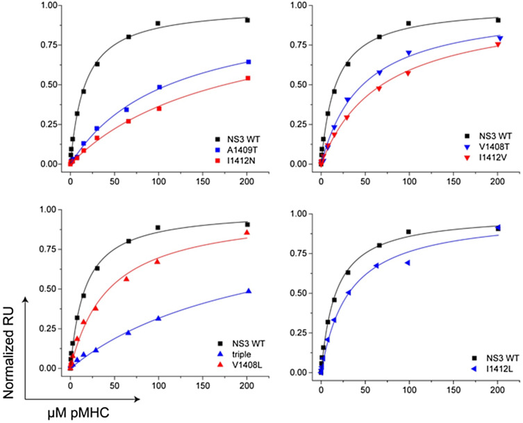
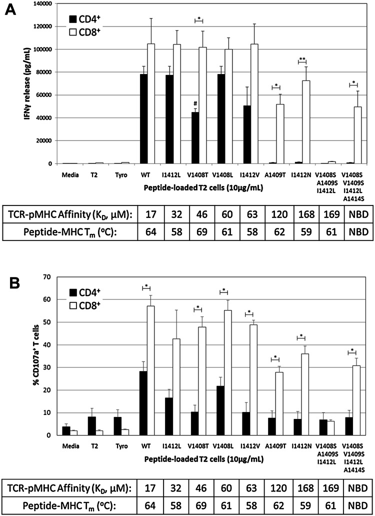
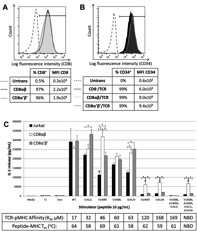
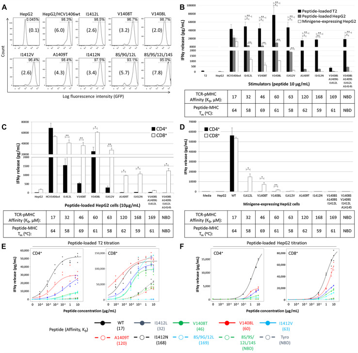
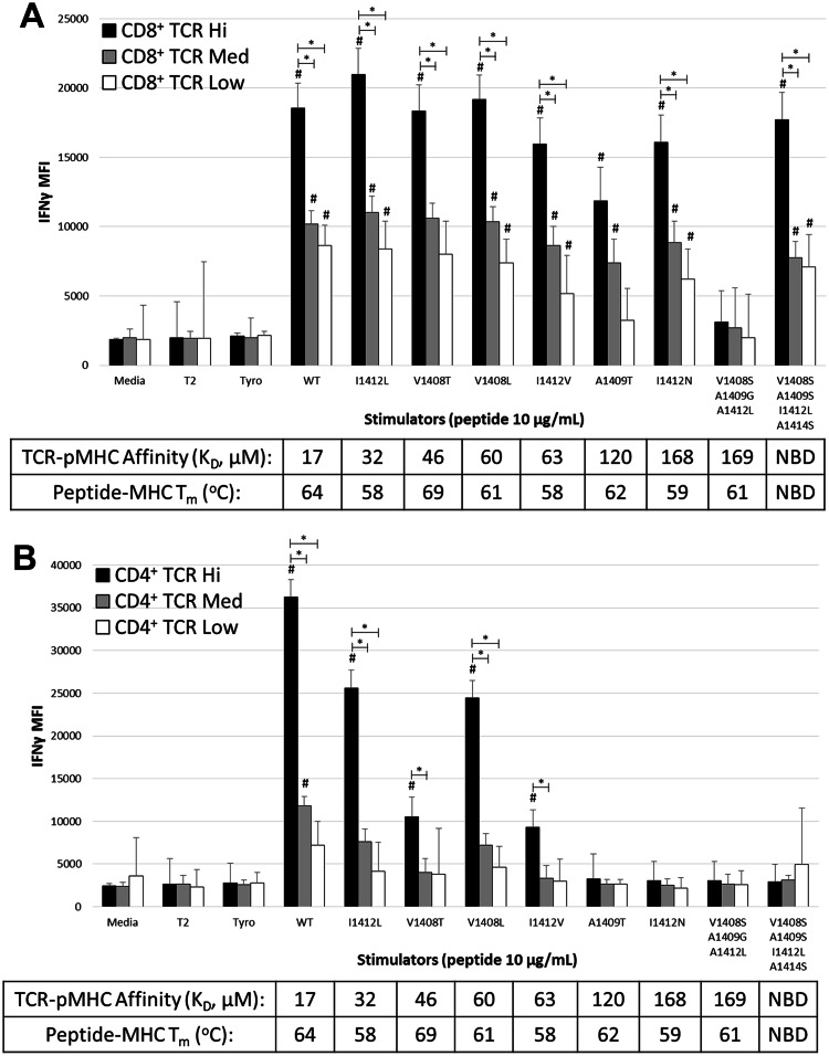
T-cell receptor (TCR)-pMHC affinity has been generally accepted to be the most important factor dictating antigen recognition in gene-modified T-cells. As such, there is great interest in optimizing TCR-based immunotherapies by enhancing TCR affinity to augment the therapeutic benefit of TCR gene-modified T-cells in cancer patients. However, recent clinical trials using affinity-enhanced TCRs in adoptive cell transfer (ACT) have observed unintended and serious adverse events, including death, attributed to unpredicted off-tumor or off-target cross-reactivity. It is critical to re-evaluate the importance of other biophysical, structural, or cellular factors that drive the reactivity of TCR gene-modified T-cells. Using a model for altered antigen recognition, we determined how TCR-pMHC affinity influenced the reactivity of hepatitis C virus (HCV) TCR gene-modified T-cells against a panel of naturally occurring HCV peptides and HCV-expressing tumor targets. The impact of other factors, such as TCR-pMHC stabilization and signaling contributions by the CD8 co-receptor, as well as antigen and TCR density were also evaluated. We found that changes in TCR-pMHC affinity did not always predict or dictate IFNγ release or degranulation by TCR gene-modified T-cells, suggesting that less emphasis might need to be placed on TCR-pMHC affinity as a means of predicting or augmenting the therapeutic potential of TCR gene-modified T-cells used in ACT. A more complete understanding of antigen recognition by gene-modified T-cells and a more rational approach to improve the design and implementation of novel TCR-based immunotherapies is necessary to enhance efficacy and maximize safety in patients.

Keywords: Adoptive cell therapy; Affinity; Altered peptide ligands; Gene-modified T-cells; T-cell; T-cell receptor (TCR).


Conflict of interest statement

The authors declare that they have no conflict of interest.

Figures

Fig. 1
Fig. 1
Surface plasmon resonance measurements of the affinity of the HCV1406 TCR for variant NS3 peptides presented by HLA-A2. Binding was measured to TCR-coated sensor surfaces by injecting increasing concentrations of pMHC. Data are shown in separate panels to facilitate comparisons; the wildtype data set (black line and black symbols) are replicated in each panel for clarity. For all measurements, data are representative of at least three separate experiments. For all mutants, data were fit using global analysis with the sensor surface as a shared parameter
Fig. 2
Fig. 2
Recognition of altered TCR–pMHC interactions is not entirely explained by changes in TCR–pMHC affinity. PBL from a normal donor were transduced with the HCV1406 TCR retroviral vector. TCR-transduced cells were enriched for CD34 expression and separated into CD4+ (black bars) and CD8+ (white bars) populations using immunomagnetic beads. T2 cells were pulsed with 10 µg/mL of WT or mutant HCV NS3:1406–1415 peptides or tyrosinase:368-376 as a control and co-culture with T-cells for 18 h. a IFNγ secretion was measured by ELISA. Mean and standard deviation of triplicate measurements of a single experiment are shown. b Degranulation of T-cells was measured by surface CD107a expression. Mean and standard deviation of three experiments for a single donor are shown. Cultures are considered “antigen-reactive” if they secrete at least 200 pg/mL IFNγ, twice above background and at least >5% of WT-stimulated cytokine release. Significant differences in IFNγ release between CD4+ and CD8+ T-cells are shown, *p < 0.05, **p < 0.01. V1408T-stimulated IFNγ release by CD4+ T-cells is significantly lower than WT, I1412L, or V1408L stimulations, # p < 0.05. SPR-measured affinities of TCR–pMHC interactions are shown, and thermal stability of peptide-MHC interactions is shown. NBD, no binding detected. Data are representative of three independent experiments with three PBL donors
Fig. 3
Fig. 3
CD8 co-receptor signaling components are required for reactivity against CD8-dependent ligands. a Jurkat cells were transduced to express either full length CD8αβ or truncated CD8α′β′ lacking the intracellular Lck-binding domain. Transduced cells were sorted for high and uniform expression of CD8. b Each group was transduced with a retrovirus encoding the HCV1406 TCR and immunomagnetically enriched for CD34 expression. c T2 cells were loaded with WT or variant HCV NS3:1406–1415 peptides or tyrosinase:368–376 peptide as a negative control and were co-cultured with TCR-transduced Jurkat cells for 18 h. IL-2 secretion by Jurkat (black bars) CD8αβ Jurkat (white bars) or CD8α′β′ (gray bars) was measured by ELISA. Mean and standard deviation of triplicate measurements are shown. Significant differences in IL-2 release between effectors are shown, *p < 0.05. Significantly less IL-2 release compared to WT stimulated is denoted with #, p < 0.05. Cultures are considered “antigen-reactive” if they secrete at least 200 pg/mL IL-2, twice above background and at least >5% of WT-stimulated cytokine release. Affinities of TCR–pMHC interactions are shown. NBD no binding detected. Data are representative of three independent experiments
Fig. 4
Fig. 4
HCV1406 TCR recognition of altered ligands is substantially diminished with lower levels of HCV antigen. a HepG2 cells were transduced with retroviral vectors encoding HCV NS3:1406–1415 variants as minigenes fused to GFP by a T2A linker. Cells were sorted for high and uniform expression of GFP. Mean fluorescence intensity (×104) is shown in parentheses for each cell line. b PBL from a normal donor were transduced with the HCV1406 TCR retroviral vector and enriched for CD34 expression using anti-CD34 immunomagnetic beads. TCR-transduced T-cells were co-cultured for 18 h with WT or mutant HCV NS3:1406–1415 peptide-loaded T2 cells (black bars), peptide-loaded HepG2 cells (gray bars) or minigene-expressing HepG2 cells (white bars). c, d HCV1406 TCR-transduced PBL were immunomagnetically isolated into CD4+ (black bars) or CD8+ (white bars) populations and co-cultured with, c HepG2 cells loaded with WT or variant HCV NS3:1406–1415 peptides or d HepG2 cells engineered to express HCV NS3:1406–1415 minigenes. IFNγ secretion after 18 h co-culture was measured by ELISA. Mean and standard deviation of triplicate measurements are shown. IFNγ secretion was measured by ELISA. Mean and standard deviation of triplicate measurements are shown. Significant differences in IFNγ release between CD4+ and CD8+ T-cells are shown, *p < 0.05, **p < 0.01. Peptide titrations (10–0.0001 μg/mL) using peptide-loaded e T2 cells or f HepG2 cells as stimulators were also performed with CD4+ (left panels) and CD8+ (right panels) TCR-transduced T-cells. Dose-dependent relationships between peptide concentration and IFNγ release were estimated using four parameter logistic models in R library ‘drc.’ EC50 estimates were extracted from the fitted model, listed in Supplemental Table S2. Cultures are considered “antigen-reactive” if they secrete at least 200 pg/mL IFNγ, twice above background and at least >5% of WT-stimulated cytokine release. Affinities of TCR–pMHC and peptide–MHC interactions are shown. SPR-measured affinities of TCR–pMHC interactions are shown, and thermal stability of peptide-MHC interactions is shown. NBD no binding detected. These data are representative of three independent experiments
Fig. 5
Fig. 5
The requirement for TCR expression to recognize altered pMHC ligands is different for CD8+ and CD4+ T-cells. PBL from a normal donor were transduced to express HCV1406 TCR and enriched for TCR-transduced cells using anti-CD34 immunomagnetic beads. T-cells were co-cultured with peptide-loaded T2 cells for 5 h and evaluated for IFNγ production by intracellular cytokine staining. Mean fluorescence intensity (MFI) of IFNγ production in TCR high (black), medium (gray), and low (white) expression populations is shown for a CD8+ and b CD4+ TCR-transduced T-cells. Significant differences in MFI of IFNγ between TCR expression levels are shown, *p < 0.05. Effector groups that display significantly greater IFNγ staining intensity compared to background tyrosinase stimulation is also denoted #p < 0.05. SPR-measured affinities of TCR–pMHC interactions are shown, and thermal stability of peptide-MHC interactions is shown. NBD no binding detected. These data are representative of three independent experiments with three donors

References

    1. Spear TT, Nagato K, Nishimura MI. Strategies to genetically engineer T cells for cancer immunotherapy. Cancer Immunol Immunother. 2016;65(6):631–649. doi: 10.1007/s00262-016-1842-5. - DOI - PMC - PubMed
    1. Tian S, Maile R, Collins EJ, Frelinger JA. CD8 + T cell activation is governed by TCR-peptide/MHC affinity, not dissociation rate. J Immunol. 2007;179(5):2952–2960. doi: 10.4049/jimmunol.179.5.2952. - DOI - PubMed
    1. Clay TM, Custer MC, Sachs J, Hwu P, Rosenberg SA, Nishimura MI. Efficient transfer of a tumor antigen-reactive TCR to human peripheral blood lymphocytes confers anti-tumor reactivity. J Immunol. 1999;163(1):507–513. - PubMed
    1. Rees W, Bender J, Teague TK, Kedl RM, Crawford F, Marrack P, Kappler J. An inverse relationship between T cell receptor affinity and antigen dose during CD4(+) T cell responses in vivo and in vitro. Proc Natl Acad Sci USA. 1999;96(17):9781–9786. doi: 10.1073/pnas.96.17.9781. - DOI - PMC - PubMed
    1. Roszkowski JJ, Lyons GE, Kast WM, Yee C, Van Besien K, Nishimura MI. Simultaneous generation of CD8 + and CD4 + melanoma-reactive T cells by retroviral-mediated transfer of a single T-cell receptor. Cancer Res. 2005;65(4):1570–1576. doi: 10.1158/0008-5472.CAN-04-2076. - DOI - PubMed

MeSH terms