

High-resolution deep functional imaging of the whole mouse brain by photoacoustic computed tomography in vivo
- PMID: 28635056
- PMCID: PMC5777675
- DOI: 10.1002/jbio.201700024
High-resolution deep functional imaging of the whole mouse brain by photoacoustic computed tomography in vivo
Abstract
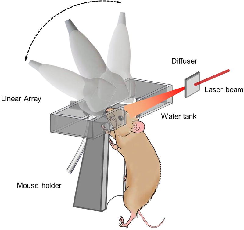
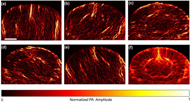
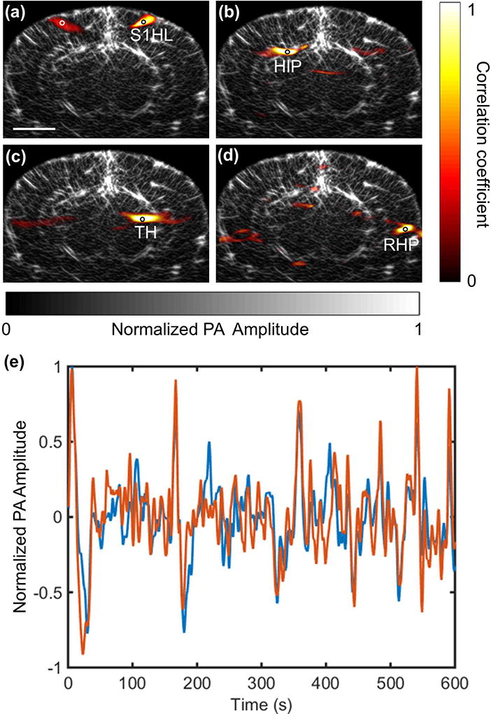
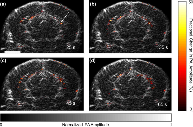
Photoacoustic computed tomography (PACT) is a non-invasive imaging technique offering high contrast, high resolution, and deep penetration in biological tissues. We report a PACT system equipped with a high frequency linear transducer array for mapping the microvascular network of a whole mouse brain with the skull intact and studying its hemodynamic activities. The linear array was scanned in the coronal plane to collect data from different angles, and full-view images were synthesized from the limited-view images in which vessels were only partially revealed. We investigated spontaneous neural activities in the deep brain by monitoring the concentration of hemoglobin in the blood vessels and observed strong interhemispherical correlations between several chosen functional regions, both in the cortical layer and in the deep regions. We also studied neural activities during an epileptic seizure and observed the epileptic wave spreading around the injection site and the wave propagating in the opposite hemisphere.
Keywords: Photoacoustic computed tomography; deep mouse brain; functional imaging.
© 2017 WILEY-VCH Verlag GmbH & Co. KGaA, Weinheim.
Figures
References
-
- Firestone LL, Gyulai F, Mintun M, Adler LJ, Urso K, Winter PM. Human brain activity response to fentanyl imaged by positron emission tomography. Anesthesia & Analgesia. 1996;82(6):1247–1251. - PubMed
-
- Macé E, Montaldo G, Cohen I, Baulac M, Fink M, Tanter M. Functional ultrasound imaging of the brain. Nature methods. 2011;8(8):662–664. - PubMed
Publication types
MeSH terms
Grants and funding
LinkOut - more resources
Full Text Sources
Other Literature Sources
Medical
Miscellaneous
