Review
. 2018 Jan 20;28(3):180-212.
doi: 10.1089/ars.2017.7120. Epub 2017 Jul 25.

Pyridine Dinucleotides from Molecules to Man

Affiliations
Review

Pyridine Dinucleotides from Molecules to Man

Joshua P Fessel et al. Antioxid Redox Signal. .

Abstract

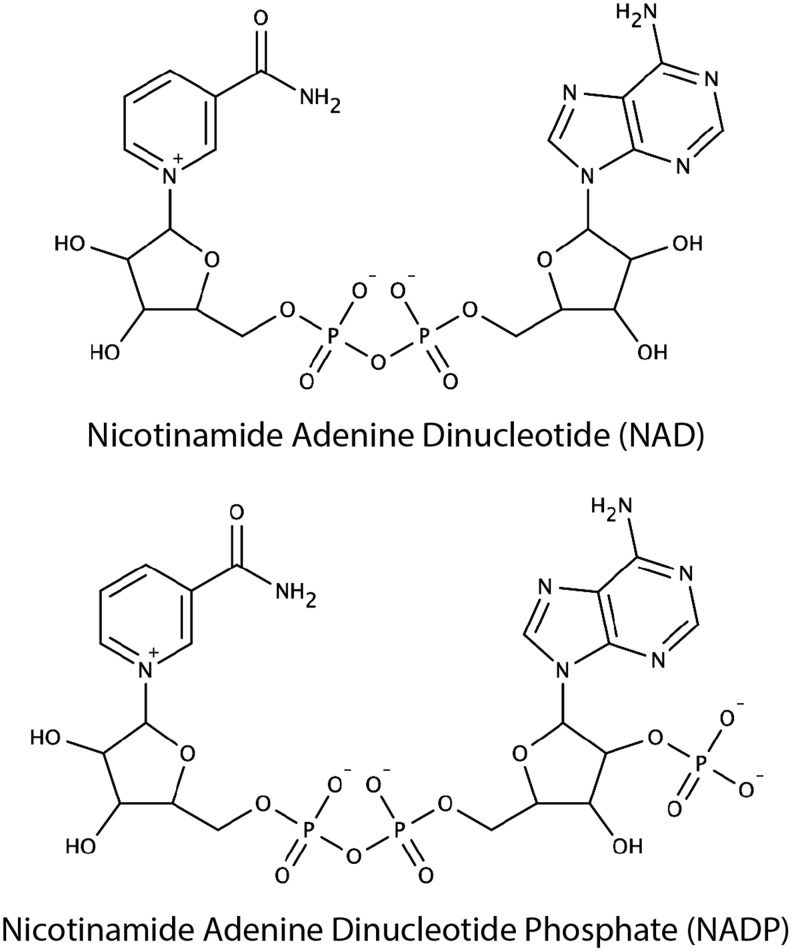
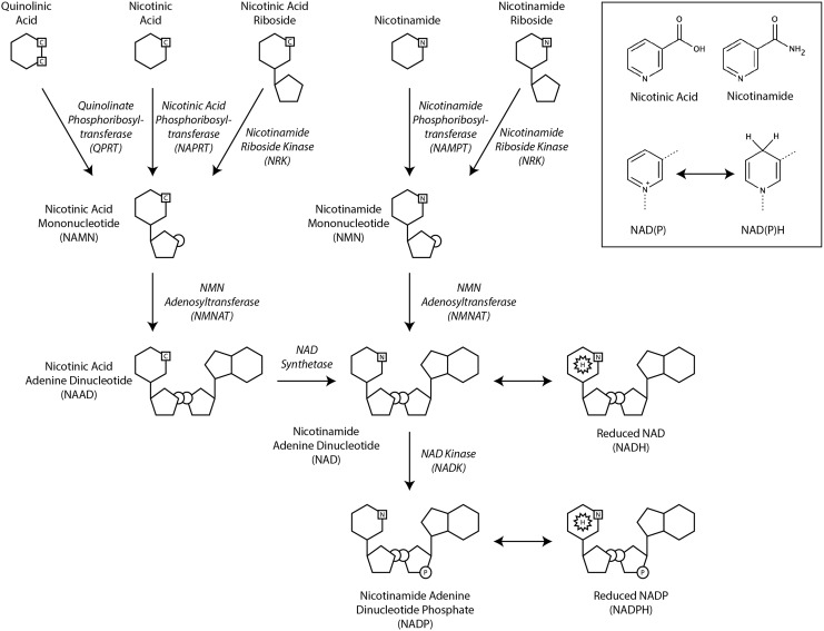
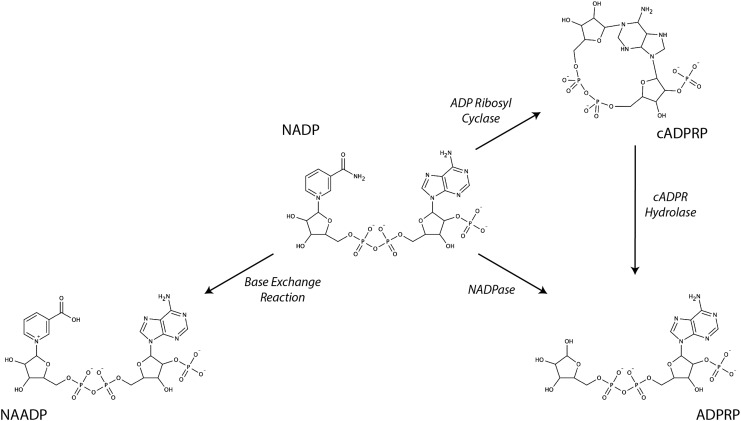
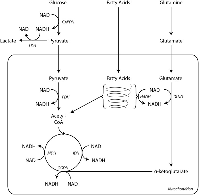
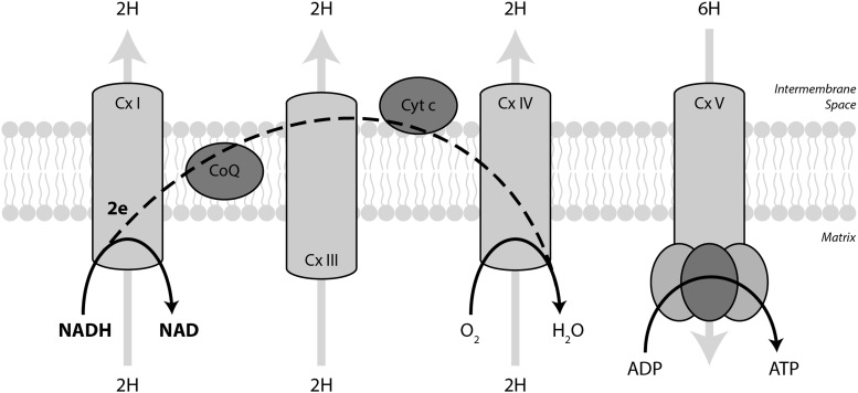
Significance: Pyridine dinucleotides, nicotinamide adenine dinucleotide (NAD) and nicotinamide adenine dinucleotide phosphate (NADP), were discovered more than 100 years ago as necessary cofactors for fermentation in yeast extracts. Since that time, these molecules have been recognized as fundamental players in a variety of cellular processes, including energy metabolism, redox homeostasis, cellular signaling, and gene transcription, among many others. Given their critical role as mediators of cellular responses to metabolic perturbations, it is unsurprising that dysregulation of NAD and NADP metabolism has been associated with the pathobiology of many chronic human diseases. Recent Advances: A biochemistry renaissance in biomedical research, with its increasing focus on the metabolic pathobiology of human disease, has reignited interest in pyridine dinucleotides, which has led to new insights into the cell biology of NAD(P) metabolism, including its cellular pharmacokinetics, biosynthesis, subcellular localization, and regulation. This review highlights these advances to illustrate the importance of NAD(P) metabolism in the molecular pathogenesis of disease.

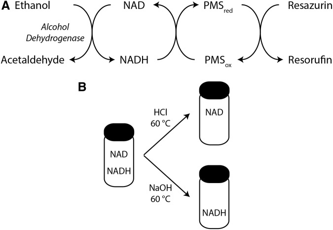
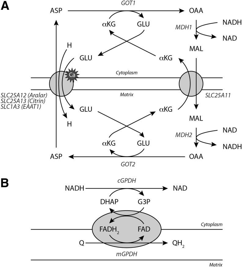
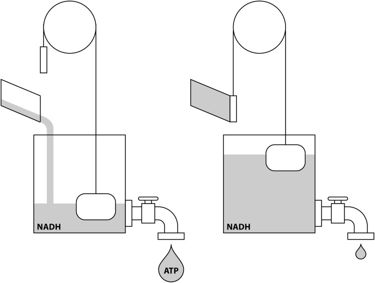
Critical issues: Perturbations of NAD(H) and NADP(H) are a prominent feature of human disease; however, fundamental questions regarding the regulation of the absolute levels of these cofactors and the key determinants of their redox ratios remain. Moreover, an integrated topological model of NAD(P) biology that combines the metabolic and other roles remains elusive.

Future directions: As the complex regulatory network of NAD(P) metabolism becomes illuminated, sophisticated new approaches to manipulating these pathways in specific organs, cells, or organelles will be developed to target the underlying pathogenic mechanisms of disease, opening doors for the next generation of redox-based, metabolism-targeted therapies. Antioxid. Redox Signal. 28, 180-212.

Keywords: NAD; NADP; PARP; SIRT; metabolism.


Figures

<b>FIG. 1.</b>
FIG. 1.
Chemical structures of NAD and NADP. Over one century ago, NAD and NADP were identified as critical factors for fermentation by yeast and named “cozymase” I and II, respectively. These nucleoside sugar phosphate molecules were subsequently found to play a key role in hydride transfer reactions during fermentation.
<b>FIG. 2.</b>
FIG. 2.
Biosynthesis of NAD and NADP. NAD is synthesized de novo from the tryptophan metabolite quinolinic acid or from the preformed precursors nicotinic acid, nicotinic acid riboside, nicotinamide, or nicotinamide riboside. The nicotinamide functional group permits the reversible electron transfer reactions that allow these pyridine dinucleotides to function as electron carriers in a variety of metabolic pathways (inset).
<b>FIG. 3.</b>
FIG. 3.
Multifunctional catalysis of NADP by CD38. CD38 catalyzes several different types of nicotinamide hydrolysis reactions using NADP as substrate. It can catalyze the cyclization of NADP to form cADPRP as well as the hydrolysis of NADP or cADPRP to form ADPRP. It can also catalyze a base exchange reaction through which nicotinamide is substituted by nicotinic acid to form NAADP. ADPRP, ADP-ribose(2′-phosphate); cADPRP, cyclic ADP-ribose(2′-phosphate); NAADP, nicotinic acid adenine dinucleotide phosphate.
<b>FIG. 4.</b>
FIG. 4.
NAD(P) half reaction. NAD(P) participates in two electron redox reactions. Note that these oxidation/reduction reactions can have consequences on cellular pH due to proton gain/loss.
<b>FIG. 5.</b>
FIG. 5.
Example of an enzymatic cycling reaction for NAD(H) quantification. (A) An example of an enzymatic cycling reaction to measure NAD(H). Electrons are transferred from ethanol via NAD and PMS to reduce resazurin to yield the fluorescent molecule resorufin at a rate dependent on the overall concentration of NAD(H). Multiple variations are possible, including the substitution of G6P and G6PD from Saccharomyces cerevisiae to measure NADP(H). Diaphorase can replace PMS, and tetrazolium salts can replace resazurin. (B) Selective degradation of NAD or NADH using buffers of different pH facilitates determination of NAD, NADH, and the NAD/NADH redox ratio from the same sample using the enzymatic cycling assay. G6P, glucose-6-phosphate; G6PD, G6P dehydrogenase; PMS, phenazine methiosulfate.
<b>FIG. 6.</b>
FIG. 6.
NADH wires cells for ATP production. All major catabolic metabolic pathways generate NADH. Glycolysis yields two NADH per glucose molecule through the activity of GAPDH. These NADH are recycled to NAD by LDH. Pyruvate is further oxidized within the mitochondria to acetyl-CoA by PDH, generating an additional molecule of NADH. During fatty acid β oxidation, HADH reduces one NAD molecule per acetyl-CoA liberated. GLUD also generates NADH during glutaminolysis. Three enzymes in the TCA cycle reduce NAD during the oxidation of substrates, IDH, OGDH complex, and MDH. Electrons from NADH then enter the electron transport chain at Complex I. ATP, adenosine triphosphate; GAPDH, glyceraldehyde 3-phosphate dehydrogenase; GLUD, glutamate dehydrogenase; HADH, 3-l-hydroxyacyl-CoA dehydrogenase; IDH, isocitrate dehydrogenase; LDH, lactate dehydrogenase; MDH, malate dehydrogenase; OGDH, α-ketoglutarate dehydrogenase; PDH, pyruvate dehydrogenase; TCA, tricarboxylic acid.
<b>FIG. 7.</b>
FIG. 7.
Shuttling NADH into the mitochondria. (A) The malate–aspartate shuttle is the main NADH translocating apparatus in mammalian cells. cMDH first transfers electrons from NADH to malate, which can enter the mitochondria via the α-ketoglutarate/malate antiporter (SLC25A11). Once inside, mMDH oxidizes malate to regenerate NADH, which can then supply electrons to Complex I. The cycle is completed by the coupled transamination of OAA to form ASP by mAST (GOT2). Aspartate leaves the mitochondria by a calcium-dependent aspartate/glutamate antiporter and is converted back to OAA by GOT1. These reactions are supported by GLU and αKG cycling. (B) NADH can also transfer electrons directly into the electron transport chain through the glycerophosphate shuttle. In this cycle, cytoplasmic GPDH oxidizes NADH to generate G3P and DHAP. G3P is then oxidized by mitochondrial GPDH thereby transferring electrons to ubiquinone via and FAD cofactor. αKG, α-ketoglutarate; ASP, aspartate; cMDH, cytoplasmic malate dehydrogenase; DHAP, dihydroxyacetone phosphate; FAD, flavin adenine dinucleotide; G3P, glycerol-3-phosphate; GLU, glutamate; GPDH, glycerol-3-phosphate dehydrogenase; mAST, mitochondrial aspartate aminotransferase; mMDH, mitochondrial malate dehydrogenase; OAA, oxaloacetate.
<b>FIG. 8.</b>
FIG. 8.
NADH supplies the electron transport chain. Mitochondrial NADH transfers its electrons to the electron transport chain through Complex I. From there, electrons travel through ubiquinone, Complex III, cytochrome c, and Complex IV where they eventually reduce molecular oxygen to water. Electron transport drives protons from the matrix to the inner membrane space to generate the chemiosmotic gradient that is coupled to ATP production by Complex V. On average, 2 electrons per NADH pumps 10 protons and yields 2.5 ATP.
<b>FIG. 9.</b>
FIG. 9.
NADH couples energy supply and demand. When energy demand is high (left panel), ATP hydrolysis and dissipation of the chemiosmotic gradient drive NADH oxidation by Complex I. When energy demand is low (right panel), accumulation of cytoplasmic and mitochondrial NADH inhibits catabolic metabolic pathways to slow bioenergetic metabolism.
<b>FIG. 10.</b>
FIG. 10.
The HIF1α transcriptional program protects against reducing stress. (A) PHD catalyzes proline hydroxylation on HIF1α in a reaction requiring O2, α-ketoglutarate, and Fe2+. (B) Hydroxylated HIF1α is recognized by the E3-ubiquitin ligase, VHL, which targets HIF1α for proteasomal degradation. As oxygen tension falls, PHD activity decreases, resulting in accumulation of unmodified HIF1α and activation of the HIF1α transcriptional program. HIF1α, hypoxia inducible factor 1α; PHD, prolyl hydroxylase; VHL, von Hippel Lindau.
<b>FIG. 11.</b>
FIG. 11.
NADPH generation by the oxidative portion of the PPP. In most cells, the PPP is the major source of NADPH. Here, G6PD consumes G6P and generates NADPH. Subsequently, 6-phosphogluconate is also oxidized to generate a second molecule of NADPH. Ribulose-5-phosphate generated from this process can then return to the glycolysis pathway as G3P and fructose-6-phosphate through the actions of transketolase and transaldolase. Alternatively, ribulose-5-phosphate can be isomerized to ribose-5-phosphate for nucleotide biosynthesis. PPP, pentose phosphate pathway.
<b>FIG. 12.</b>
FIG. 12.
NADPH provides electrons for nucleotide biosynthesis. RNR, which catalyzes dehydroxylation of NDP to dNDP, forms an active site disulfide during catalysis that must be reduced for continued enzymatic activity. Electrons are transferred from NADPH through two redox pathways. In one, GR oxidizes NADPH to reduce GSSG to GSH. Glutathione reduces disulfides in Grx that subsequently catalyzes the reduction of disulfides in RNR. In the other pathway, NADPH is utilized by TrxR to reduce disulfides in Trx, which, in turn, reduces disulfides in RNR. dNDP, deoxyribonucleotide diphosphates; GR, glutathione reductase; Grx, glutaredoxin; GSH, glutathione; GSSG, glutathione disulfide; NDP, ribonucleotide diphosphates; RNR, ribonucleotide reductase; Trx, thioredoxin; TrxR, thioredoxin reductase.
<b>FIG. 13.</b>
FIG. 13.
NADPH provides electrons to enzymes involved in antioxidant defense. A variety of redox transfer reactions utilize NADPH as the initial electron donor to reduce and detoxify ROS. GSH generated by glutathione reductase is consumed by Gpx enzymes. Thioredoxin also participates in a variety of chemical reductions designed to mitigate oxidant stress through reduction of Prx, H2O2, and other proteins. Gpx, glutathione peroxidase; Prx, peroxiredoxin; ROS, reactive oxygen species.
<b>FIG. 14.</b>
FIG. 14.
The polyol pathway. Under conditions of significant glucose excess, hexokinase, the initial enzyme in glycolysis, becomes saturated. The accumulating glucose is diverted into the polyol pathway where glucose is metabolized to sorbitol by aldose reductase and sorbitol is metabolized to fructose by sorbitol dehydrogenase. The first reaction requires NADPH and the second reaction yields NADH. Thus, these reactions yield a net exchange of NADPH for NADH, which is likely deleterious under conditions of reductive stress (e.g., diabetes-induced increases in glycolytic flux lowering the NAD/NADH ratio leading to ROS production in the setting of impaired antioxidant defense due to NADPH deficiency).

References

    1. Institute of Medicine. Dietary Reference Intakes for Thiamin, Riboflavin, Niacin, Vitamin B6, Folate, Vitamin B12, Pantothenic Acid, Biotin, and Choline. Washington, DC: The National Academies Press; 1998, p. 592 - PubMed
    1. Alano CC, Tran A, Tao R, Ying W, Karliner JS, and Swanson RA. Differences among cell types in NAD(+) compartmentalization: a comparison of neurons, astrocytes, and cardiac myocytes. J Neurosci Res 85: 3378–3385, 2007 - PubMed
    1. Ali RA, Camick C, Wiles K, Walseth TF, Slama JT, Bhattacharya S, Giovannucci DR, and Wall KA. Nicotinic acid adenine dinucleotide phosphate plays a critical role in naive and effector murine T cells but not natural regulatory T cells. J Biol Chem 291: 4503–4522, 2016 - PMC - PubMed
    1. Anderson KA, Green MF, Huynh FK, Wagner GR, and Hirschey MD. SnapShot: mammalian sirtuins. Cell 159: 956–956.e1, 2014 - PMC - PubMed
    1. Avalos JL, Bever KM, and Wolberger C. Mechanism of sirtuin inhibition by nicotinamide: altering the NAD(+) cosubstrate specificity of a Sir2 enzyme. Mol Cell 17: 855–868, 2005 - PubMed

MeSH terms

LinkOut - more resources