. 2017 Jun 21;18(6):1332.
doi: 10.3390/ijms18061332.

The Application of Heptamethine Cyanine Dye DZ-1 and Indocyanine Green for Imaging and Targeting in Xenograft Models of Hepatocellular Carcinoma

Affiliations

The Application of Heptamethine Cyanine Dye DZ-1 and Indocyanine Green for Imaging and Targeting in Xenograft Models of Hepatocellular Carcinoma

Caiqin Zhang et al. Int J Mol Sci. .

Abstract

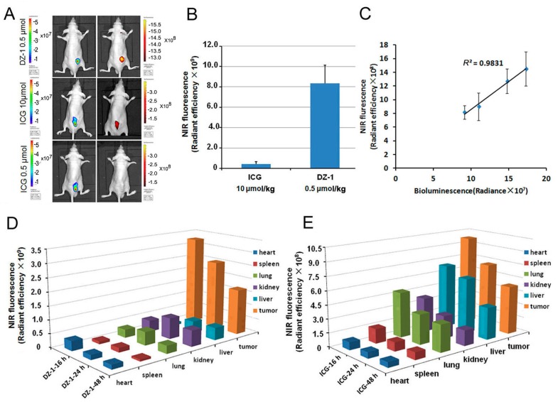
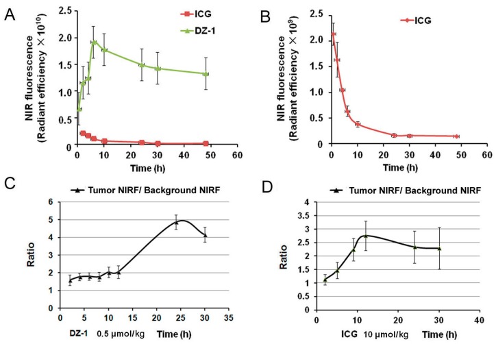
Near infrared fluorescence (NIRF) imaging has strong potential for widespread use in noninvasive tumor imaging. Indocyanine green (ICG) is the only Food and Drug Administration (FDA) -approved NIRF dye for clinical diagnosis; however, it is unstable and poorly targets tumors. DZ-1 is a novel heptamethine cyanine NIRF dye, suitable for imaging and tumor targeting. Here, we compared the fluorescence intensity and metabolism of DZ-1 and ICG. Additionally, we assayed their specificities and abilities to target tumor cells, using cultured hepatocellular carcinoma (HCC) cell lines, a nude mouse subcutaneous xenograft model of liver cancer, and a rabbit orthotopic transplantation model. We found that DZ-1 accumulates in tumor tissue and specifically recognizes HCC in subcutaneous and orthotopic models. The NIRF intensity of DZ-1 was one order of magnitude stronger than that of ICG, and DZ-1 showed excellent intraoperative tumor targeting in the rabbit model. Importantly, ICG accumulated at tumor sites, as well as in the liver and kidney. Furthermore, DZ-1 analog-gemcitabine conjugate (NIRG) exhibited similar tumor-specific targeting and imaging properties, including inhibition of tumor growth, in HCC patient-derived xenograft (PDX) mice. DZ-1 and NIRG demonstrated superior tumor-targeting specificity, compared to ICG. We show that DZ-1 is an effective molecular probe for specific imaging, targeting, and therapy in HCC.

Keywords: hepatocellular carcinoma; heptamethine cyanine dyes; intraoperative navigation; near-infrared fluorescence; optical imaging; xenograft model.


Conflict of interest statement

The authors declare no conflict of interest.

Figures

Figure 1
Figure 1
Uptake of DZ-1 and indocyanine green (ICG) in hepatocellular carcinoma (HCC). (A) Confocal microscope analyses of DZ-1 dye uptake by HCC. Original magnification: 400×; scale bars: 50 μm; (B) Ratio of near infrared fluorescence (NIRF, red) dye uptake intensity in HCC at successive time points. Data are presented as mean ± standard deviation (SD) (n = 5); (C,D) Co-localization of DZ-1 or ICG (red) and LysoTracker (green, top panel) or MitoTracker (yellow, bottom panel) in Hep3B cells, as determined by confocal microscopy. Cell nuclei were stained with 4′,6-diamidino-2-phenylindole (DAPI, blue). Original magnification: 1200×; Scale bars, 20 μm.
Figure 1
Figure 1
Uptake of DZ-1 and indocyanine green (ICG) in hepatocellular carcinoma (HCC). (A) Confocal microscope analyses of DZ-1 dye uptake by HCC. Original magnification: 400×; scale bars: 50 μm; (B) Ratio of near infrared fluorescence (NIRF, red) dye uptake intensity in HCC at successive time points. Data are presented as mean ± standard deviation (SD) (n = 5); (C,D) Co-localization of DZ-1 or ICG (red) and LysoTracker (green, top panel) or MitoTracker (yellow, bottom panel) in Hep3B cells, as determined by confocal microscopy. Cell nuclei were stained with 4′,6-diamidino-2-phenylindole (DAPI, blue). Original magnification: 1200×; Scale bars, 20 μm.
Figure 2
Figure 2
The distribution of DZ-1 and ICG in the organs of subcutaneous tumor model. (A) Dual ex vivo BLI/NIRF imaging of mice with Hep3B-Luc subcutaneous xenografts treated with varying doses of ICG or DZ-1; (B) Quantification of NIRF intensity within the tumor area (per cm2) of subcutaneous tumor xenografts; (C) NIRF/BLI signal intensity correlation in mice (n = 5) with subcutaneous tumor xenografts (right); (D,E) Distribution intensity per cm2 of DZ-1 and ICG in the organs of subcutaneous tumor models at successive times.
Figure 3
Figure 3
Metabolism of DZ-1 and ICG in the organs of subcutaneous tumor model. (A,B) NIRF intensities per unit area (cm2) in subcutaneous tumor xenograft mice subjected to DZ-1 or ICG were measured at 0.5, 2, 3, 6, 10, 24, and 48 h; (C,D). Ratio of tumor NIRF intensity to background NIRF intensity at various time points after injection of DZ-1 or ICG in subcutaneous tumor xenograft mice.
Figure 4
Figure 4
Comparison of ICG and DZ-1 imaging in orthotopic liver tumors. (A) Representative NIRF optical images of tumor sites by injection of DZ-1 (0.5 μmol/kg) in vivo. Scale bar: 20 μm. (B) Representative NIRF optical images of tumor sites by injection of ICG (10 μmol/kg) in vivo. However, there were more differences between the bioluminescence signal range and fluorescence in the tumor site in vitro. Scale bar: 20 μm. (C) A large area of carcinogenesis of liver identified by ICG (10 μmol/kg). The ex vivo signal range of bioluminescence in the organ is consistent with that of fluorescence.
Figure 5
Figure 5
The application of DZ-1 and ICG in surgical exploration. (A) DZ-1 imaging using NIRF optical fiber intraoperative guidance in rabbits with VX2 liver cancer (left). Ex vivo NIRF imaging of rabbit liver cancer using small animal optical imaging system (right). Hematoxylin-eosin (H&E) staining of the fluorescent tissue sections; (B) NIRF intensity/tumor area (per cm2) of DZ-1 uptake in liver tumor of rabbit; (C) ICG imaging using NIRF optical fiber intraoperative guidance in rabbits with VX2 liver cancer (left). Ex vivo NIRF imaging of rabbit liver cancer using small animal optical imaging system (right). H&E staining of the fluorescent tissue sections. 10× and 20×, Scale bars, 20 μm; (D) NIRF intensity/tumor area (per cm2) of ICG uptake in liver tumor of the rabbits. Blue circle indicate the tumor location.
Figure 6
Figure 6
Imaging and targeting of DZ-1 drug conjugate in a liver cancer patient-derived xenograft (PDX) model. (A) H&E, NIRF, and immunohistochemistry (IHC) analyses of liver cancer tissues derived from both PDX mouse models and original patient samples. Original magnification: 400×; scale bars represent 20 μm; (B) DZ-1 dye uptake by Hep3B cells with a prior exposure to either HIF1α stabilizers (DMOG), or OATP inhibitor (BSP). Scale bar, 50 μm; (C) NIRF optical imaging of PDX models established by implanting 3 different human liver cancer specimens to subcutaneous of nude mice. Strong fluorescent signal was detected at subcutaneous tumor site; (DF) Inhibition of NIRG on the tumor growth from liver cancer PDX mouse, including C64003, C34566, and B66873. d represents the treatment time.

Similar articles

Cited by

References

    1. Reinier H., Sun H., England C.G., Valdovinos H.F., Ehlerding E.B., Barnhart T.E., Yang Y., Cai W. CD146-targeted immunoPET and NIRF imaging of hepatocellular carcinoma with a dual-labeled monoclonal antibody. Theranostics. 2016;6:1918–1933. - PMC - PubMed
    1. Yang G., Yi Z., Li X., Jie L., Wang J.P., Chordia M.D., Zhong Q.L., Chung L.W.K., Wei Y., Pan D. Improving therapeutic potential of farnesylthiosalicylic acid: Tumor specific delivery via conjugation with heptamethine cyanine dye. Mol. Pharm. 2016;14:1–13. - PMC - PubMed
    1. Trivedi E.R., Harney A.S., Olive M.B., Podgorski I., Moin K., Sloane B.F., Barrett A.G., Meade T.J., Hoffman B.M. Chiral porphyrazine near-IR optical imaging agent exhibiting preferential tumor accumulation. Proc. Natl. Acad. Sci. USA. 2010;107:1284–1288. doi: 10.1073/pnas.0912811107. - DOI - PMC - PubMed
    1. Frangioni J.V. In vivo near-infrared fluorescence imaging. Curr. Opin. Chem. Biol. 2003;7:626–634. doi: 10.1016/j.cbpa.2003.08.007. - DOI - PubMed
    1. Shi C., Wu J.B., Pan D. Review on near-infrared heptamethine cyanine dyes as theranostic agents for tumor imaging, targeting, and photodynamic therapy. J. Biomed. Opt. 2016;21:50901. doi: 10.1117/1.JBO.21.5.050901. - DOI - PubMed

MeSH terms

LinkOut - more resources