Review
. 2017 Jun 21;9(6):69.
doi: 10.3390/cancers9060069.

Selection of Nucleic Acid Aptamers Targeting Tumor Cell-Surface Protein Biomarkers

Affiliations
Review

Selection of Nucleic Acid Aptamers Targeting Tumor Cell-Surface Protein Biomarkers

Marie-Cécile Mercier et al. Cancers (Basel). .

Abstract

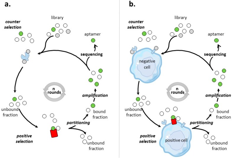
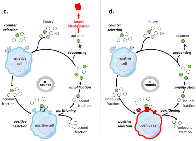
Aptamers are nucleic acids referred to as chemical antibodies as they bind to their specific targets with high affinity and selectivity. They are selected via an iterative process known as 'selective evolution of ligands by exponential enrichment' (SELEX). Aptamers have been developed against numerous cancer targets and among them, many tumor cell-membrane protein biomarkers. The identification of aptamers targeting cell-surface proteins has mainly been performed by two different strategies: protein- and cell-based SELEX, when the targets used for selection were proteins and cells, respectively. This review aims to update the literature on aptamers targeting tumor cell surface protein biomarkers, highlighting potentials, pitfalls of protein- and cell-based selection processes and applications of such selected molecules. Aptamers as promising agents for diagnosis and therapeutic approaches in oncology are documented, as well as aptamers in clinical development.

Keywords: SELEX; aptamer; cell-surface biomarker.


Conflict of interest statement

The authors declare no conflict of interest.

Figures

Figure 1
Figure 1
Scheme of protein- and cell-based SELEX processes. Briefly, the process of SELEX involves first a selection step: the nucleic acid library is incubated with a target (positive selection), which can be preceded or followed by a counter selection phase to remove non-specific nucleic acid molecules. During the partitioning step, bound and unbound fractions are separated. The bound fraction is amplified to obtain an enriched pool for next round of selection. This process is repeated for n rounds until the pool is enriched for sequences that specifically bind the target. These nucleic acid molecules are cloned and sequenced. Individual sequences are aptamers. (a) Protein-based SELEX. The pre-identified purified protein used as target for the protein-based SELEX is colored in red. (b) Cell-based SELEX on a pre-identified tumor cell-surface biomarker colored in red. (c) Cell-based SELEX on a post-identified tumor cell-surface biomarker. The target, identified at the end of the SELEX process, is colored in red. (d) Cell-based SELEX to a tumor cell type. In this case, no particular cell-surface biomarkers are identified and aptamers are specific to the cell’s molecular signature. The whole cell used for positive selection is colored in red.
Figure 1
Figure 1
Scheme of protein- and cell-based SELEX processes. Briefly, the process of SELEX involves first a selection step: the nucleic acid library is incubated with a target (positive selection), which can be preceded or followed by a counter selection phase to remove non-specific nucleic acid molecules. During the partitioning step, bound and unbound fractions are separated. The bound fraction is amplified to obtain an enriched pool for next round of selection. This process is repeated for n rounds until the pool is enriched for sequences that specifically bind the target. These nucleic acid molecules are cloned and sequenced. Individual sequences are aptamers. (a) Protein-based SELEX. The pre-identified purified protein used as target for the protein-based SELEX is colored in red. (b) Cell-based SELEX on a pre-identified tumor cell-surface biomarker colored in red. (c) Cell-based SELEX on a post-identified tumor cell-surface biomarker. The target, identified at the end of the SELEX process, is colored in red. (d) Cell-based SELEX to a tumor cell type. In this case, no particular cell-surface biomarkers are identified and aptamers are specific to the cell’s molecular signature. The whole cell used for positive selection is colored in red.

References

    1. Ellington A.D., Szostak J.W. In vitro selection of RNA molecules that bind specific ligands. Nature. 1990;346:818–822. doi: 10.1038/346818a0. - DOI - PubMed
    1. Robertson D.L., Joyce G.F. Selection in vitro of an RNA enzyme that specifically cleaves single-stranded DNA. Nature. 1990;344:467–468. doi: 10.1038/344467a0. - DOI - PubMed
    1. Tuerk C., Gold L. Systematic evolution of ligands by exponential enrichment: RNA ligands to bacteriophage T4 DNA polymerase. Science. 1990;249:505–510. doi: 10.1126/science.2200121. - DOI - PubMed
    1. Ponomarenko J.V., Orlova G.V., Frolov A.S., Gelfand M.S., Ponomarenko M.P. SELEX_DB: A database on in vitro selected oligomers adapted for recognizing natural sites and for analyzing both SNPs and site-directed mutagenesis data. Nucleic Acids Res. 2002;30:195–199. doi: 10.1093/nar/30.1.195. - DOI - PMC - PubMed
    1. Aptamer Database Apta-Index™. [(accessed on 1 June 2017)]; Available online: http://www.aptagen.com/aptamer-index/aptamer-list.aspx.