. 2017 Jun 20;112(12):2567-2574.
doi: 10.1016/j.bpj.2017.05.006.

Heterogeneous Tau-Tubulin Complexes Accelerate Microtubule Polymerization

Affiliations

Heterogeneous Tau-Tubulin Complexes Accelerate Microtubule Polymerization

Xiao-Han Li et al. Biophys J. .

Abstract

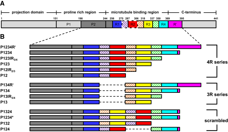
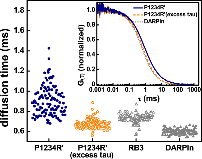
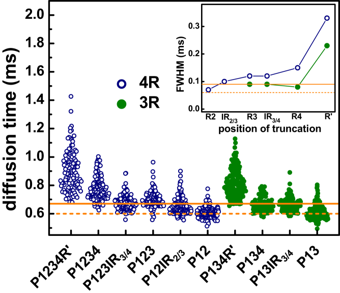
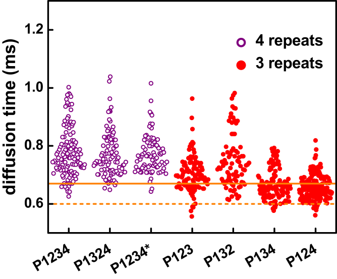
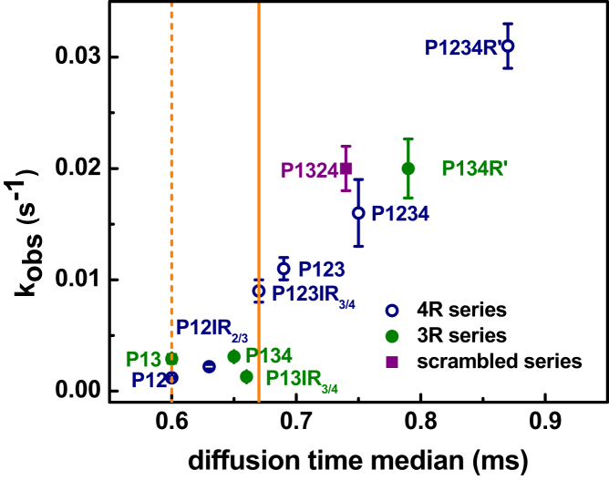
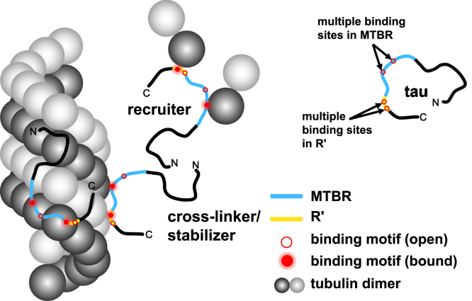
Tau is an intrinsically disordered protein with a central role in the pathology of a number of neurodegenerative diseases. Tau normally functions to stabilize neuronal microtubules, although the mechanism underlying this function is not well understood. Of note is that the interaction between tau and soluble tubulin, which has implications both in understanding tau function as well as its role in disease, is underexplored. Here we investigate the relationship between heterogeneity in tau-tubulin complexes and tau function. Specifically, we created a series of truncated and scrambled tau constructs and characterized the size and heterogeneity of the tau-tubulin complexes formed under nonpolymerizing conditions. Function of the constructs was verified by tubulin polymerization assays. We find that, surprisingly, the pseudo-repeat region of tau, which flanks the core microtubule-binding domain of tau, contributes largely to the formation of large, heterogeneous tau tubulin complexes; additional independent tubulin binding sites exist in repeats two and three of the microtubule binding domain. Of particular interest is that we find positive correlation between the size and heterogeneity of the complexes and rate of tau-promoted microtubule polymerization. We propose that tau-tubulin can be described as a "fuzzy" complex, and our results demonstrate the importance of heterogeneous complex formation in tau function. This work provides fundamental insights into the functional mechanism of tau, and more broadly underscores the relevance of heterogeneous and dynamic complexes in the functions of intrinsically disordered proteins.


Figures

Figure 1
Figure 1
Schematics of tau constructs. (A) Given here is the longest isoform of human tau. The regions of interest are color-coded: P2, dark gray; R1, blue; IR1/2, purple; R2, red; IR2/3, orange; R3, yellow; IR3/4, green; R4, cyan; and R′, magenta. The inter-repeats are indicated by hatch marks. IR1/2–R2, highlighted by dashed lines, is alternatively spliced to generate 3R/4R tau. The numbers on top of the schematic indicate residues delineating both major domains and subdomains of interest to this study. (B) Given here are the constructs used in this work. The color-code for each construct matches that in (A). Gray dashed lines indicate deletion of specific subdomains.
Figure 2
Figure 2
Heterogeneity of tau-tubulin complexes. Shown here is the distribution of the diffusion times derived from fits of individual autocorrelation curves. (Inset) Given here are normalized average autocorrelation curves of P1234R′-tubulin with excess tubulin or excess tau and DARPin-tubulin. To see this figure in color, go online.
Figure 3
Figure 3
Quantification of heterogeneity of tau-tubulin complexes. Diffusion times of tau-tubulin complexes for constructs based on truncation of 4R and 3R tau are plotted. The lines at 0.60 and 0.67 ms denote the upper (solid) and lower (dashed) boundaries of expected median diffusion times of 1:1 tau-tubulin complexes determined by FCS measurements with excess P1234R′ and P13, respectively, as described in the text. (Inset) The widths of Gaussian fits of each distribution of diffusion times are shown. The lines are full width half-maximum from the measurements corresponding to the lines of median diffusion times in the main plot. Details are in the text and Supporting Material. To see this figure in color, go online.
Figure 4
Figure 4
Tubulin binding sites in R2 and R3. Diffusion times are plotted of tau-tubulin complexes formed by tau constructs containing either four or three microtubule binding repeats. The solid and dashed lines denote the upper and lower boundaries, respectively, of 1:1 tau-tubulin complexes as described in the text and in Fig. 3. The four-repeat series, in which all constructs contain both IR1/2–R2 and IR2/3–R3, bind with a >1:1 stoichiometry and significant heterogeneity. In the three-repeat series, only P123 and P132 bind with >1:1 stoichiometry. Constructs lacking either IR1/2–R2 (P134) or IR2/3–R3 (P124) have smaller median diffusion times, reflecting smaller complexes and lower stoichiometry. To see this figure in color, go online.
Figure 5
Figure 5
Correlation between diffusion times and polymerization rates. The inverse of polymerization half-time (kobs) for each construct is plotted against the median diffusion time for each construct. The error bars indicate standard deviations in the rate of polymerization. The solid and dashed lines denote the upper and lower boundaries, respectively, for the median diffusion time of 1:1 tau-tubulin determined using labeled tubulin as described in the text and Supporting Material. To see this figure in color, go online.
Figure 6
Figure 6
Model of tau-tubulin fuzzy complexes. Tau contains multiple tubulin binding motifs (red circles) located in the MTBR (blue) and R′ (yellow). These motifs bind to tubulin or microtubules stochastically and dynamically. This mode of interaction allows tau to perform functional roles as a stabilizer, cross-linker, and recruiter. Variable combinations of binding motifs work cooperatively to stabilize microtubule and promote microtubule assembly.

References

    1. Ballatore C., Lee V.M.Y., Trojanowski J.Q. Tau-mediated neurodegeneration in Alzheimer’s disease and related disorders. Nat. Rev. Neurosci. 2007;8:663–672. - PubMed
    1. Mazanetz M.P., Fischer P.M. Untangling tau hyperphosphorylation in drug design for neurodegenerative diseases. Nat. Rev. Drug Discov. 2007;6:464–479. - PubMed
    1. Weingarten M.D., Lockwood A.H., Kirschner M.W. A protein factor essential for microtubule assembly. Proc. Natl. Acad. Sci. USA. 1975;72:1858–1862. - PMC - PubMed
    1. Drubin D.G., Kirschner M.W. Tau protein function in living cells. J. Cell Biol. 1986;103:2739–2746. - PMC - PubMed
    1. Trojanowski J.Q., Lee V.M.Y. Pathological tau: a loss of normal function or a gain in toxicity? Nat. Neurosci. 2005;8:1136–1137. - PubMed

LinkOut - more resources