Review
. 2017 Jun 20;46(6):910-926.
doi: 10.1016/j.immuni.2017.05.011.

The Enteric Network: Interactions between the Immune and Nervous Systems of the Gut

Affiliations
Review

The Enteric Network: Interactions between the Immune and Nervous Systems of the Gut

Bryan B Yoo et al. Immunity. .

Abstract

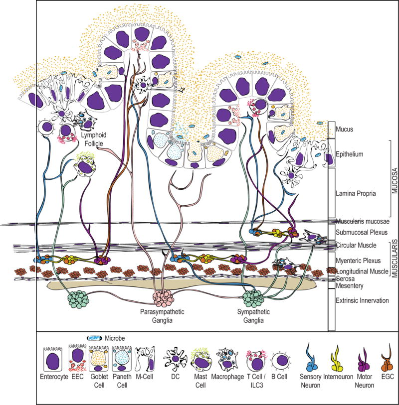
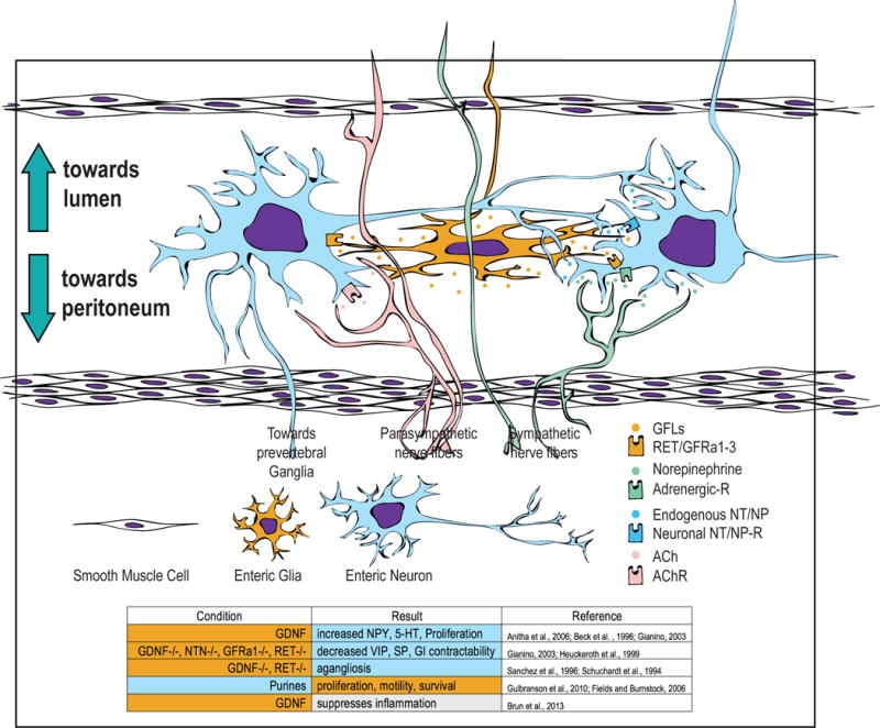
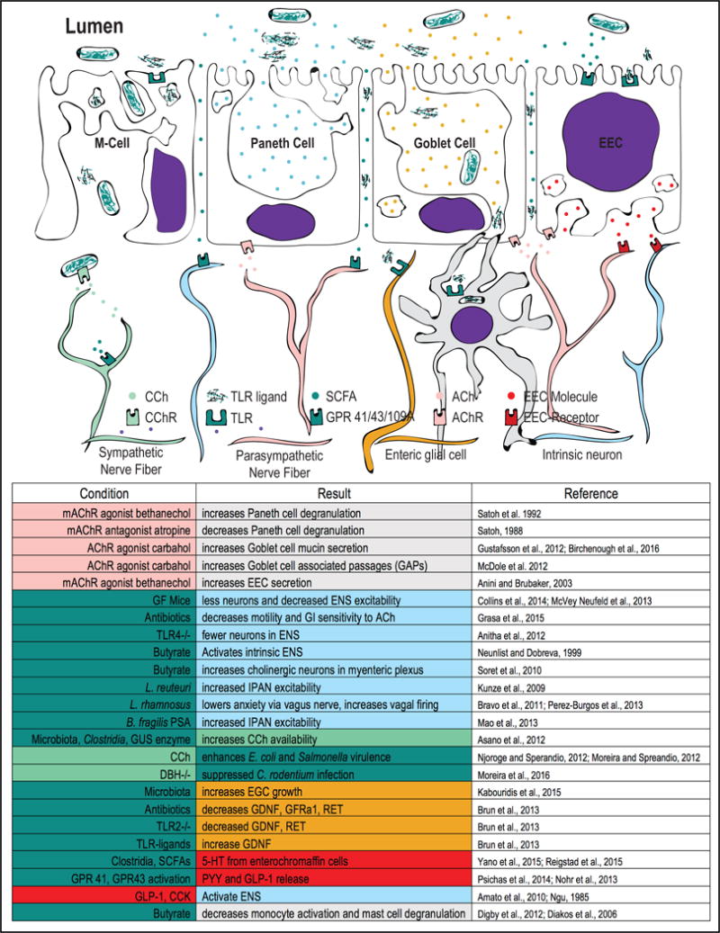
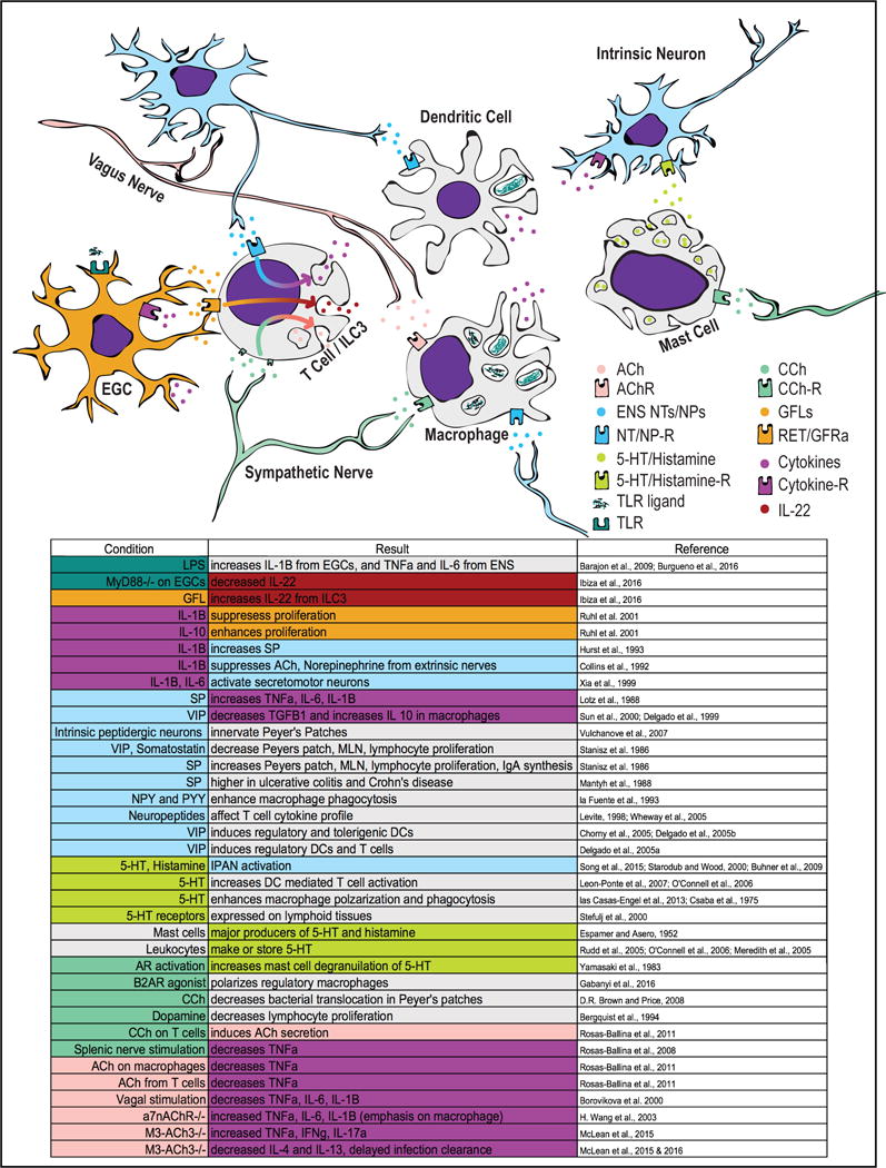
Interactions between the nervous and immune systems enable the gut to respond to the variety of dietary products that it absorbs, the broad spectrum of pathogens that it encounters, and the diverse microbiome that it harbors. The enteric nervous system (ENS) senses and reacts to the dynamic ecosystem of the gastrointestinal (GI) tract by translating chemical cues from the environment into neuronal impulses that propagate throughout the gut and into other organs in the body, including the central nervous system (CNS). This review will describe the current understanding of the anatomy and physiology of the GI tract by focusing on the ENS and the mucosal immune system. We highlight emerging literature that the ENS is essential for important aspects of microbe-induced immune responses in the gut. Although most basic and applied research in neuroscience has focused on the brain, the proximity of the ENS to the immune system and its interface with the external environment suggest that novel paradigms for nervous system function await discovery.

Keywords: enteric nervous system; gastrointestinal tract; gut-brain axis; intestinal microbiota; neuro-immune interactions; neuro-immunity.


Figures

Figure 1
Figure 1. Anatomy of the GI Tract
The GI tract is comprised of distinct cross-sectional compartments. Extrinsic, sympathetic and parasympathetic nerve fibers enter the GI tract through the mesentery and can extend throughout all layers of intestinal tissue. The myenteric and submucosal plexuses form the dense nerve network that innervates the entire length and depth of the GI tract. Various immune cells are resident to the muscularis, but are also highly abundant in the lamina propria, especially in Peyer’s patches and lymphoid follicles. These immune cells are also in close proximity to neurons and glia. The epithelium shown here is made up of 5 different cell types. These include absorptive enterocytes, EECs, goblet cells, Paneth cells, and M-cells.
Figure 2
Figure 2. Connectivity of Enteric Neurons and Glia
Enteric neurons are located in either the submucosal or myenteric plexuses. Both plexuses are located in between two muscle layers (see Figure 1). Parasympathetic nerve fibers release acetylcholine and sympathetic nerves release norepinephrine. These extrinsic nerve fibers can innervate enteric neurons but also associate with the cells in the smooth muscle, lamina propria, and the epithelium. Enteric neurons can innervate one another or extend towards the lamina propria, and specific intestinofugal (IFANs) can synapse onto sympathetic ganglia. Enteric glial cells make and release neurotrophic factors, associate with enteric neurons, and extend throughout the mucosa. The left and middle columns are color coded to represent cells and molecules that generate specific conditions and the results that are produced from those conditions, respectively.
Figure 3
Figure 3. Interactions at the Intestinal Epithelium
The intestinal epithelium is where luminal constituents are actively or passively transported into the tissue. Extrinsic nerves and neurons are found near the epithelium, and thus the molecules that cross the epithelium and those that are secreted basolaterally can potentially have an effect on their activity. Microbes or microbial parts can cross the epithelium and affect other cell types through M-cells, immunoglobulin-mediated transcytosis, goblet cell-associated passages (GAPs), and by general leakiness of the epithelium. Dendritic cells (DCs) and macrophages can phagocytose microbial antigens and secrete cytokines that can have an effect on neurons as well. Parasympathetic fibers release acetylcholine (ACh) and induce secretion of intracellular stores of molecules. In goblet cells, ACh also increases rates of DC luminal sampling via GAPs. EECs can also release neuroendocrine molecules in response to TLR stimulation and SCFAs. These molecules released basolaterally can potentially regulate the activity of neurons. Enteric glial cells can also project towards the epithelium, potentially allowing microbes to impact their function. Enteric neurons can be activated by commensal and pathogenic bacteria, as well as short chain fatty acids that diffuse across the epithelium. The left and middle columns are color coded to represent cells and molecules that generate specific conditions and the results that are produced from those conditions, respectively.
Figure 4
Figure 4. Interactions between GI Immune Cells and the ENS
Extrinsic nerves, intrinsic neurons, and enteric glial cells are in close proximity to each other and to the immune cells in the GI tract. Thus, the molecules that are produced by one cell can have an effect on another cell, given that the latter expresses a receptor to recognize the molecule. By these parameters, interactions between neurons/glia and immune cells are, in theory, abundant, and some of these putative interactions are presented here. Immune cells can be influenced by neurotransmitters and neuropeptides produced by intrinsic neurons of the ENS (as well as those produced by extrinsic nerve fibers), and cytokines produced by immune cells can have a reciprocal effect on neurons. The left and middle columns are color coded to represent cells and molecules that generate specific conditions and the results that are produced from those conditions, respectively.

References

    1. Aas J, Gessert CE, Bakken JS. Recurrent Clostridium difficile colitis: case series involving 18 patients treated with donor stool administered via a nasogastric tube. Clin Infect Dis. 2003;36:580–585. doi: 10.1086/367657. - DOI - PubMed
    1. Abad C, Martinez C, Juarranz MG, Arranz A, Leceta J, Delgado M, Gomariz RP. Therapeutic effects of vasoactive intestinal peptide in the trinitrobenzene sulfonic acid mice model of Crohn’s disease. Gastroenterology. 2003;124:961–971. doi: 10.1053/gast.2003.50141. - DOI - PubMed
    1. Ahlman H, Lundberg J, Dahlstrom A, Kewenter J. A Possible Vagal Adrenergic Release of Serotonin from Enterochromaffin Cells in the Cat. Acta Physiologica. 1976;98:366–375. doi: 10.1111/j.1748-1716.1976.tb10321.x. - DOI - PubMed
    1. Ahmad A, Wang CH, Bell RG. A role for IgE in intestinal immunity. Expression of rapid expulsion of Trichinella spiralis in rats transfused with IgE and thoracic duct lymphocytes. J Immunol. 1991;146:3563–3570. - PubMed
    1. Amato A, Cinci L, Rotondo A, Serio R, Faussone pellegrini MS, Vannucchi MG, Mule F. Peripheral motor action of glucagon-like peptide-1 through enteric neuronal receptors. Neurogastroenterol Motil. 2010;22:664–e203. doi: 10.1111/j.1365-2982.2010.01476.x. - DOI - PubMed

Publication types