. 2017 Aug 21;45(14):8378-8391.
doi: 10.1093/nar/gkx521.

Functional and structural analysis of AT-specific minor groove binders that disrupt DNA-protein interactions and cause disintegration of the Trypanosoma brucei kinetoplast

Affiliations

Functional and structural analysis of AT-specific minor groove binders that disrupt DNA-protein interactions and cause disintegration of the Trypanosoma brucei kinetoplast

Cinthia R Millan et al. Nucleic Acids Res. .

Abstract

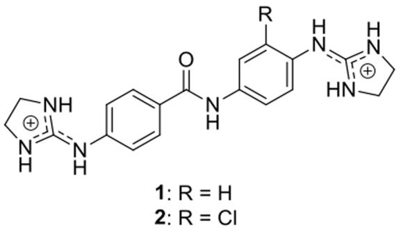
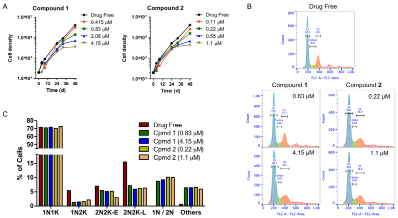
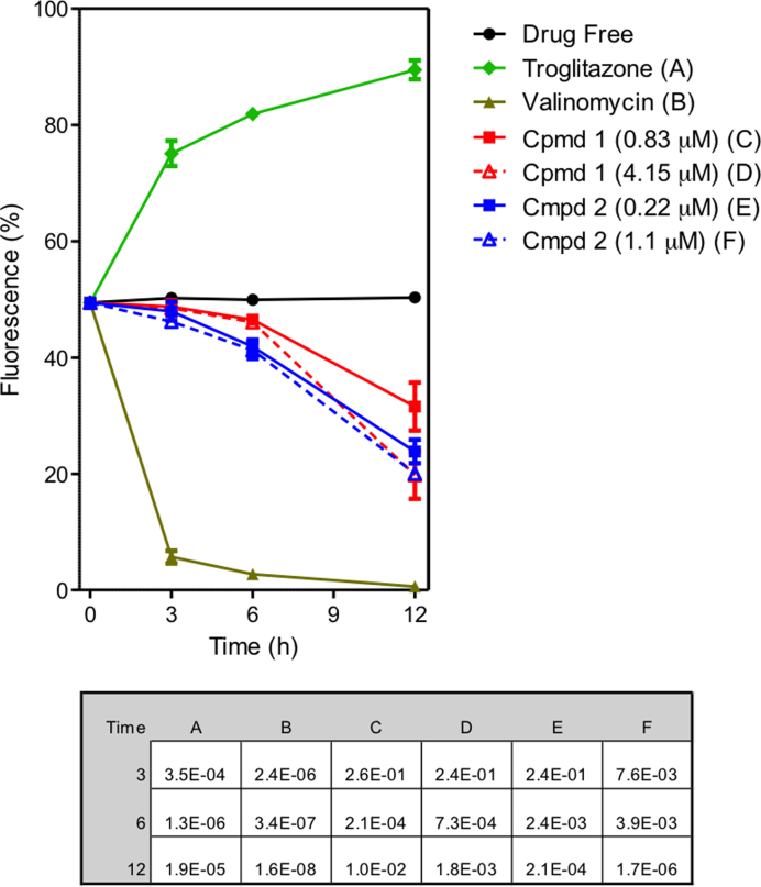
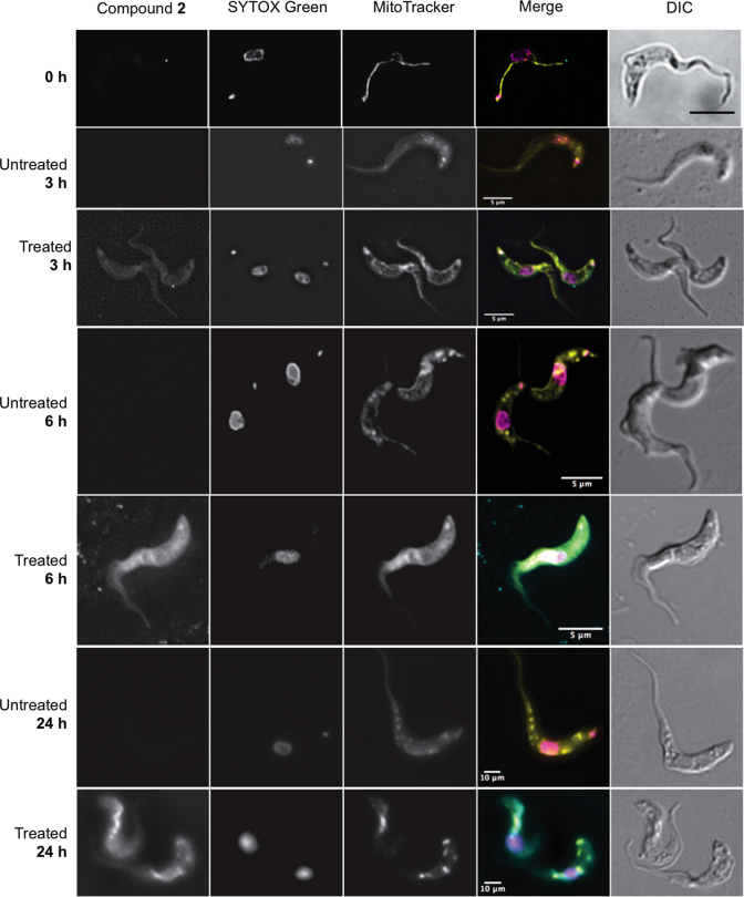
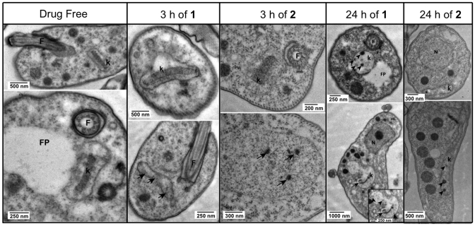
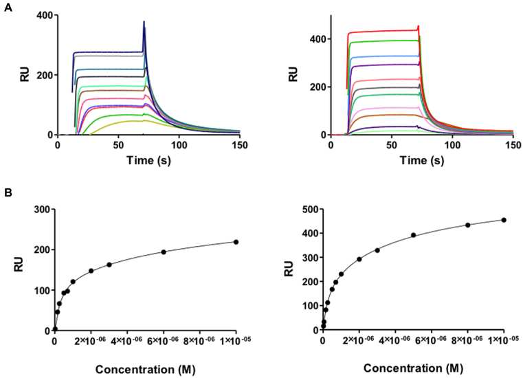
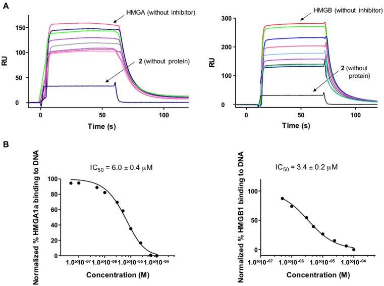
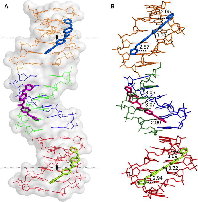
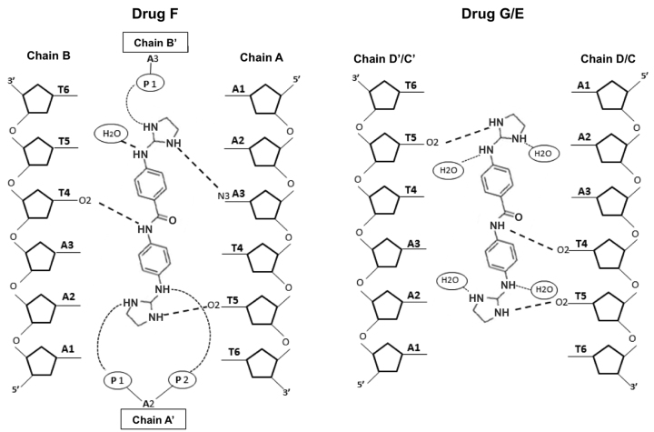
Trypanosoma brucei, the causative agent of sleeping sickness (Human African Trypanosomiasis, HAT), contains a kinetoplast with the mitochondrial DNA (kDNA), comprising of >70% AT base pairs. This has prompted studies of drugs interacting with AT-rich DNA, such as the N-phenylbenzamide bis(2-aminoimidazoline) derivatives 1 [4-((4,5-dihydro-1H-imidazol-2-yl)amino)-N-(4-((4,5-dihydro-1H-imidazol-2-yl)amino)phenyl)benzamide dihydrochloride] and 2 [N-(3-chloro-4-((4,5-dihydro-1H-imidazol-2-yl)amino)phenyl)-4-((4,5-dihydro-1H-imidazol-2-yl)amino)benzamide] as potential drugs for HAT. Both compounds show in vitro effects against T. brucei and in vivo curative activity in a mouse model of HAT. The main objective was to identify their cellular target inside the parasite. We were able to demonstrate that the compounds have a clear effect on the S-phase of T. brucei cell cycle by inflicting specific damage on the kinetoplast. Surface plasmon resonance (SPR)-biosensor experiments show that the drug can displace HMG box-containing proteins essential for kDNA function from their kDNA binding sites. The crystal structure of the complex of the oligonucleotide d[AAATTT]2 with compound 1 solved at 1.25 Å (PDB-ID: 5LIT) shows that the drug covers the minor groove of DNA, displaces bound water and interacts with neighbouring DNA molecules as a cross-linking agent. We conclude that 1 and 2 are powerful trypanocides that act directly on the kinetoplast, a structure unique to the order Kinetoplastida.


Figures

Figure 1.
Figure 1.
Structure of compounds 1 and 2.
Figure 2.
Figure 2.
(A) Growth curves of untreated control T. brucei 427WT and of parallel cultures treated with compounds 1 and 2 at 0.5, 1, 2 and 5 × their EC50 values. (B) Histograms of flow cytometric analysis of propidium iodide fluorescence associated with Tb427WT trypanosomes. The frames shown show control and exposed (1 × and 5 × EC50) to 1 or 2 for 24 h, with most cells in G1 phase (blue peak, single set of chromosomes) and smaller numbers in S-phase (green, DNA synthesis) and G2 phase (orange, 2 sets of chromosomes). (C) DNA content of cells treated 24 h with compounds 1 and 2 at 1 × and 5 × EC50 as determined by fluorescence microscopy (N, nucleus; K, kinetoplast; 1N/2N, cells with one or two nuclei but no observable kinetoplastid).
Figure 3.
Figure 3.
Effect of compounds 1 and 2 on mitochondrial membrane potential (Ψm) of T. b. brucei s427-WT. The results shown are the mean of three independent determinations; error bars depict standard errors. The inset table lists the P values, using a Student's two-tailed, unpaired t-test comparing each category to the untreated control, incubated for the same length of time; a value <0.05 is considered to be significant. Untreated cells (drug free), valinomycin (negative - depolarization) and troglitazone (positive - hyperpolarization) are employed as controls.
Figure 4.
Figure 4.
Fluorescence localization of the compound 2 in Trypanosoma brucei s427-WT cells. Images were taken at 3, 6 and 24 h of incubation with 5 μM compound 2. Untreated cell samples were taken at each time point for drug free control. All fluorescent images are shown with compound 2 (λ = 450, blue channel), SYTOX Green (λ = 523, green channel), MitoTracker (λ = 599, red channel), and merge, where arbitrary colors were used to visualize the various dyes: blue for 2, purple for SYTOX, yellow for Mitotracker. The outline of all cells is shown by differential interference contrast (DIC) imaging. Images were acquired using a DeltaVision imaging system and deconvolved using the ratio conservative method, on SoftWoRx software.
Figure 5.
Figure 5.
TEM images showing normal ultrastructure of bloodstream form for treated and untreated (drug free control) cells of Tb427-WT after incubation with compound 1 or compound 2 for either 3 h or 24 h. F = flagellum, FP = flagellum pocket, k = kinetoplast. Images were observed in a Tecnai T20 (FEI) at 200 kV. Irregular structures in the kinetoplast are shown with arrows and arrowheads, indicating kDNA damage.
Figure 6.
Figure 6.
(A) SPR sensorgrams of HMGA1a (top left panel) and HMGB1 (top right panel) binding to dsDNA biotin-GGGGAATAATCGCGATTATTCCCCAATAATCGCGATTATT in HEPES 1 at 25°C. For HMGA1a, the best sensorgrams were obtained in HEPES 1 buffer, instead of MES or Phosphate buffer (43). (B) Binding curves for interaction of HMGA1a (lower left panel) and HMGB1 (lower right panel) with target DNA and fitting curve for a two-site affinity model.
Figure 7.
Figure 7.
(A) SPR competition sensorgrams showing the inhibition of a fixed concentration (2 μM) of HMGA1a (Top left panel) and HMGB1 (top right panel) binding to dsDNA containing AATAAT_ATTATT oligonucleotide in the presence of increasing concentration of 2. Concentration range from 0.05 to 200 μM (for HMGA1a) and 0.05 to 400 μM (for HMGB1). (B) Inhibition curves and IC50 values for inhibition of binding of HMGA1a (lower left panel) and HMGB1 (lower right panel) to dsDNA by compound 2.
Figure 8.
Figure 8.
(A) View of the different crystallographic units of the complex. The black lozenge indicates the dyad axes. There are four independent single oligonucleotides chains; two of them (blue-green) form the central duplex and the other two (orange and red) form two different DNA duplexes with their symmetrical chain. Three crystallographically independent drug molecules are indicated in different colours. Drug F (pink), Drugs E (green) and G (blue). (B) Hydrogen bonds formed by the drugs with the minor-groove atoms of the DNA duplexes show similar interactions. The orientation of the aromatic rings in the central drug (F, pink) differs from to the other two drugs. Drug E and G have two possible inverted positions in the groove; for clarity only one of them is shown.
Figure 9.
Figure 9.
Schematic representation of interactions between F and G or E drugs and d[AAATTT]2. In the crystal, drugs G and E may be found in two alternative positions, up and down, which are structurally identical. Symmetric chains are indicated with apostrophe.

References

    1. World Health Organization and Department of control of neglected tropical diseases Report of the second WHO stakeholders meeting on gambiense human African trypanosomiasis elimination. 2016; Geneva: 21–23.
    1. Delespaux V., de Koning H.P.. Drugs and drug resistance in African trypanosomiasis. Drug Resist. Updat. 2007; 10:30–50. - PubMed
    1. Ríos Martínez C.H., Nué Martínez J.J., Ebiloma G.U., de Koning H.P., Alkorta I., Dardonville C.. Lowering the pKa of a bisimidazoline lead with halogen atoms results in improved activity and selectivity against Trypanosoma brucei in vitro. Eur. J. Med. Chem. 2015; 101:806–817. - PubMed
    1. Ríos Martínez C.H., Miller F., Ganeshamoorthy K., Glacial F., Kaiser M., de Koning H.P., Eze A.A., Lagartera L., Herraiz T., Dardonville C.. A new nonpolar N-hydroxy imidazoline lead compound with improved activity in a murine model of late-stage Trypanosomabrucei brucei infection is not cross-resistant with diamidines. Antimicrob. Agents Chemother. 2015; 59:890–904. - PMC - PubMed
    1. Dardonville C., Barrett M.P., Brun R., Kaiser M., Tanious F., Wilson W.D.. DNA binding affinity of bisguanidine and bis(2-aminoimidazoline) derivatives with in vivo antitrypanosomal activity. J. Med. Chem. 2006; 49:3748–3752. - PubMed

MeSH terms