. 2017 Jun 21;12(1):11.
doi: 10.1186/s13064-017-0088-z.

The emergence of mesencephalic trigeminal neurons

Affiliations

The emergence of mesencephalic trigeminal neurons

Marcela Lipovsek et al. Neural Dev. .

Abstract

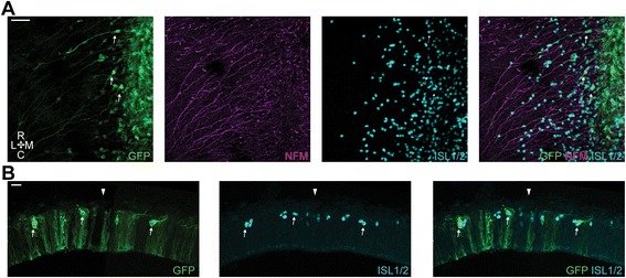
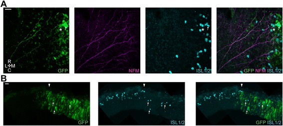
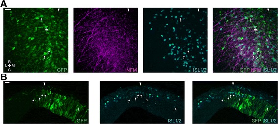
Background: The cells of the mesencephalic trigeminal nucleus (MTN) are the proprioceptive sensory neurons that innervate the jaw closing muscles. These cells differentiate close to the two key signalling centres that influence the dorsal midbrain, the isthmus, which mediates its effects via FGF and WNT signalling and the roof plate, which is a major source of BMP signalling as well as WNT signalling.

Methods: In this study, we have set out to analyse the importance of FGF, WNT and BMP signalling for the development of the MTN. We have employed pharmacological inhibitors of these pathways in explant cultures as well as utilising the electroporation of inhibitory constructs in vivo in the chick embryo.

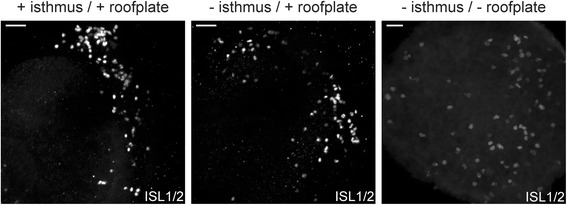
Results: We find that interfering with either FGF or WNT signalling has pronounced effects on MTN development whilst abrogation of BMP signalling has no effect. We show that treatment of explants with either FGF or WNT antagonists results in the generation of fewer MTN neurons and affects MTN axon extension and that inhibition of both these pathways has an additive effect. To complement these studies, we have used in vivo electroporation to inhibit BMP, FGF and WNT signalling within dorsal midbrain cells prior to, and during, their differentiation as MTN neurons. Again, we find that inhibition of BMP signalling has no effect on the development of MTN neurons. We additionally find that cells electroporated with inhibitory constructs for either FGF or WNT signalling can differentiate as MTN neurons suggesting that these pathways are not required cell intrinsically for the emergence of these neurons. Indeed, we also show that explants of dorsal mesencephalon lacking both the isthmus and roof plate can generate MTN neurons. However, we did find that inhibiting FGF or WNT signalling had consequences for MTN differentiation.

Conclusions: Our results suggest that the emergence of MTN neurons is an intrinsic property of the dorsal mesencephalon of gnathostomes, and that this population undergoes expansion, and maturation, along with the rest of the dorsal midbrain under the influence of FGF and WNT signalling.

Keywords: FGF; Jaw proprioception; MTN; MesV; Mesencephalic trigeminal nucleus; Midbrain; WNT BMP.


Figures

Fig. 1
Fig. 1
Pharmacological inhibition of signalling pathways on stage 13 explant cultures. a. Wholemount dorsal view of explant cultures treated with, from left to right, DMSO, dorsomorphin, SU5402, IWP-2 and SU5402 + IWP-2 and immunostained for NFM and ISL1/2. Filled arrowheads show the position of the dorsal midline. Empty arrowheads show the position of the trigeminal placode. R, rostral. C, caudal. Scale bars, 50 μm. Insets, Wholemount dorsal view of explant cultures treated with DMSO or dorsomorphin and immunostained for phospho-SMAD1/5/8 and ISL1/2. Scale bars, 50 μm. b. Bar graphs showing ISL1/2+ cell counting on stage 13 explants. Values are mean ± S.E.M. normalised to the mean control value of each experiment. *, significant p values for SU5402 and IWP-2 treatments after multiple comparison using Kruskal-Wallis test with Dunn’s correction and for the SU5402 + IWP-2 for pairwise comparison using two-tailed Mann Whitney test. Dorsomorphin: DMSO (n = 39), 5 μM (n = 19), 10 μM (n = 13). SU5402: DMSO (n = 33), 34 μM (n = 12), 68 μM (n = 16). IWP-2: DMSO (n = 24), 1 μM (n = 10), 5 μM (n = 16). SU5402 + IWP-2: DMSO (n = 9), 68 μM + 5 μM (n = 9). Replicates are from >3independent experiments
Fig. 2
Fig. 2
Pharmacological inhibition of signalling pathways on stage 11 explant cultures. a. Wholemount dorsal view of explant cultures treated with, from left to right, DMSO, dorsomorphin, SU5402, IWP-2 and SU5402 + IWP-2 and immunostained for NFM and ISL1/2. Filled arrowheads show the position of the dorsal midline. Empty arrowheads show the position of the trigeminal placode. R, rostral. C, caudal. Scale bars, 50 μm. b. Bar graphs showing ISL1/2+ cell counting on stage 13 explants. Values are mean ± S.E.M. normalised to the mean control value of each experiment. *, significant p values for pairwise comparisons using two-tailed Mann Whitney test. Dorsomorphin: DMSO (n = 9), 10 μM (n = 12). SU5402: DMSO (n = 12), 68 μM (n = 9). IWP-2: DMSO (n = 9), 5 μM (n = 14). SU5402 + IWP-2: DMSO (n = 9), 68 μM + 5 μM (n = 7). Replicates are from >3 independent experiments
Fig. 3
Fig. 3
In ovo electroporation of the dorsal midbrain. a. Representative wholemount dorsal view of a midbrain electroporated with CAGGS-GFP (n = 25). Arrowhead denotes the position of the dorsal midline. Thin arrows, typical MTN neurons. Big arrow, right side LLF. Scale bar, 50 μm. b. Higher magnification image of GFP-electroporated cells. Thin arrows, typical MTN neurons. Scale bar, 20 μm. c. Transverse cryosection of a GFP electroporated midbrain. Arrowhead denotes the position of the dorsal midline. Thin arrows, typical MTN neurons. Scale bar, 20 μm. R, rostral. C, caudal. M, medial. L, lateral
Fig. 4
Fig. 4
Inhibition of BMP signalling via in ovo electroporation of the dorsal midbrain. a. Representative high magnification image of Smad6-IRES-GFP electroporated cells. Thin arrows, typical MTN neurons. Scale bar, 25 μm (n = 8). b. Transverse cryosection of a Smad6-IRES-GFP electroporated midbrain. Arrowhead denotes the position of the dorsal midline. Thin arrows, typical MTN neurons. Scale bar, 20 μm
Fig. 5
Fig. 5
Inhibition of FGF signalling via in ovo electroporation of the dorsal midbrain. a, b, c and d. Representative high magnification images of dnFGFR1-IRES-GFP electroporated cells illustrating the different morphologies observed (n = 18). Thin arrows, typical dnFGFR1 electroporated MTN neurons. Scale bars, 20 μm. e. Transverse cryosection of a dnFGFR1-IRES-GFP electroporated midbrain. Arrowhead denotes the position of the dorsal midline. Thin arrows, typical dnFGFR1 electroporated MTN neurons. Scale bar, 20 μm
Fig. 6
Fig. 6
Inhibition of WNT signalling via in ovo electroporation of the dorsal midbrain. a. Representative high magnification image of GSK3-IRES-GFP electroporated cells (n = 19). Thin arrow, typical GSK3 electroporated MTN neuron. Asterisk, deeper MTN neuron. Scale bar, 20 μm. b. Transverse cryosection of a GKS3-IRES-GFP electroporated midbrain. Arrowhead denotes the position of the dorsal midline. Thin arrows, typical GSK3 electroporated MTN neurons. Scale bar, 20 μm
Fig. 7
Fig. 7
Inhibition of FGF and WNT signalling via in ovo electroporation of the dorsal midbrain. a. Representative high magnification image of dnFGFR1-IRES-GFP and GSK3-IRES-GFP electroporated cells (n = 11). Thin arrow, typical dnFGFR1 + GSK3 electroporated MTN neurons. Asterisk, Stubby GFP+ cell. Arrowhead denotes the position of the dorsal midline. Scale bar, 20 μm. b. Transverse cryosection of a dnFGFR1-IRES-GFP and GKS3-IRES-GFP electroporated midbrain. Arrowhead denotes the position of the dorsal midline. Thin arrows, typical GSK3 electroporated MTN neurons. Scale bar, 20 μm
Fig. 8
Fig. 8
Cultured explants of midbrain neural tube. Representative wholemount views of midbrain explant cultures. Left, whole midbrain with the isthmus (n = 6); middle, midbrain without the isthmus (n = 22); right midbrain without the isthmus and roof plate (n = 38). Immunostained for ISL1/2. Scale bars. 50 μm

References

    1. DAMICOMARTEL A, NODEN D. Contributions of PLACODAL and neural crest cells to avian cranial peripheral ganglia. Am J Anat. 1983;166(4):445–468. doi: 10.1002/aja.1001660406. - DOI - PubMed
    1. Begbie J, Ballivet M, Graham A. Early steps in the production of sensory neurons by the neurogenic placodes. Mol Cell Neurosci. 2002;21(3):502–511. doi: 10.1006/mcne.2002.1197. - DOI - PubMed
    1. Harlow D, Barlow L. Embryonic origin of gustatory cranial sensory neurons. Dev Biol. 2007;310(2):317–328. doi: 10.1016/j.ydbio.2007.07.042. - DOI - PMC - PubMed
    1. O'Neill P, Mak SS, Fritzsch B, Ladher RK, Baker CV. The amniote paratympanic organ develops from a previously undiscovered sensory placode. Nat Commun. 2012;3:1041. doi: 10.1038/ncomms2036. - DOI - PMC - PubMed
    1. Thompson H, Blentic A, Watson S, Begbie J, Graham A. The formation of the superior and jugular ganglia: insights into the generation of sensory neurons by the neural crest. Dev Dyn. 2010;239(2):439–445. doi: 10.1002/dvdy.22179. - DOI - PubMed

LinkOut - more resources