

Cell Death in the Vessel Wall: The Good, the Bad, the Ugly
- PMID: 28637702
- PMCID: PMC5584709
- DOI: 10.1161/ATVBAHA.117.309229
Cell Death in the Vessel Wall: The Good, the Bad, the Ugly
Abstract
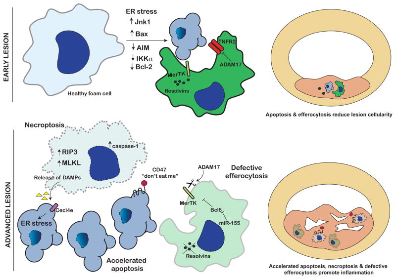
Atherosclerotic vascular disease results from an imbalance of inflammatory and vascular cell accumulation and removal in the neointimal space. When pathways that promote cell recruitment, survival and proliferation are favored over to those that activate cell death, egress and clearance, the plaque expands. In contrast, programmed cell death and the efficient clearance of apoptotic bodies by efferocytosis reduces lesion cellularity and promotes a reparative environment and lesion stability. However, should these carefully balanced pathways become disturbed, lesions can accumulate cell debris, damaged-associated molecular patterns and arrested macrophages, all contributing to the pro-inflammatory environment and lesion instability. Here, we will review the latest understanding of how cell death in the vessel wall directly coordinates the development of atherosclerosis, and what molecular signals are orchestrating these pathways. We will discuss the necessity of cell death, and the ways in which the execution of different forms of cell death can direct different outcomes in the plaque, and how promoting the effective clearance of dead cells from the lesion is looking like a promising therapeutic path forward.
Keywords: apoptosis; atherosclerosis; cell death; macrophage; necrosis.
Figures
References
-
- Kockx MM, Herman AG. Apoptosis in atherosclerosis: beneficial or detrimental? Cardiovascular research. 2000;45:736–46. - PubMed
-
- Arai S, Shelton JM, Chen M, Bradley MN, Castrillo A, Bookout AL, Mak PA, Edwards PA, Mangelsdorf DJ, Tontonoz P, Miyazaki T. A role for the apoptosis inhibitory factor AIM/Spalpha/Api6 in atherosclerosis development. Cell Metab. 2005;1:201–13. - PubMed
-
- Li Y, Schwabe RF, DeVries-Seimon T, Yao PM, Gerbod-Giannone MC, Tall AR, Davis RJ, Flavell R, Brenner DA, Tabas I. Free cholesterol-loaded macrophages are an abundant source of tumor necrosis factor-alpha and interleukin-6: model of NF-kappaB- and map kinase-dependent inflammation in advanced atherosclerosis. J Biol Chem. 2005;280:21763–72. - PubMed
Publication types
MeSH terms
Substances
Grants and funding
LinkOut - more resources
Full Text Sources
Other Literature Sources
Medical
Research Materials
