. 2017 Aug 10;91(17):e00539-17.
doi: 10.1128/JVI.00539-17. Print 2017 Sep 1.

Identification of an Intermediate in Hepatitis B Virus Covalently Closed Circular (CCC) DNA Formation and Sensitive and Selective CCC DNA Detection

Affiliations

Identification of an Intermediate in Hepatitis B Virus Covalently Closed Circular (CCC) DNA Formation and Sensitive and Selective CCC DNA Detection

Jun Luo et al. J Virol. .

Abstract

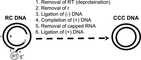
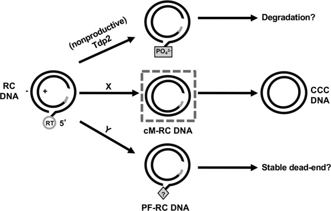
Hepatitis B virus (HBV) covalently closed circular (CCC) DNA functions as the only viral template capable of coding for all the viral RNA species and is thus essential to initiate and sustain viral replication. CCC DNA is converted, in a multistep and ill-understood process, from a relaxed circular (RC) DNA, in which neither of the two DNA strands is covalently closed. To detect putative intermediates during RC DNA to CCC DNA conversion, two 3' exonucleases, exonuclease I (Exo I) and Exo III, were used in combination to degrade all DNA strands with a free 3' end, which would nevertheless preserve closed circular DNA in either single-stranded (SS) or double-stranded (DS) form. Indeed, an RC DNA species with a covalently closed minus strand but an open plus strand (closed minus-strand RC DNA [cM-RC DNA]) was detected by this approach. Further analyses indicated that at least some of the plus strands in such a putative intermediate likely still retained the RNA primer that is attached to the 5' end of the plus strand in RC DNA, suggesting that minus-strand closing can occur before plus-strand processing. Furthermore, the same nuclease treatment proved to be useful for sensitive and specific detection of CCC DNA by removing all DNA species other than closed circular DNA. Application of these and similar approaches may allow the identification of additional intermediates during CCC DNA formation and facilitate specific and sensitive detection of CCC DNA, which should help elucidate the pathways of CCC DNA formation and the factors involved.IMPORTANCE The hepatitis B virus (HBV) covalently closed circular (CCC) DNA, by serving as the viral transcriptional template, is the molecular basis of viral persistence. CCC DNA is converted, in a multistep and ill-understood process, from relaxed circular (RC) DNA. Little is currently understood about the pathways or factors involved in CCC DNA formation. We have now detected a likely intermediate during the conversion of RC DNA to CCC DNA, thus providing important clues to the pathways of CCC DNA formation. Furthermore, the same experimental approach that led to the detection of the intermediate could also facilitate specific and sensitive detection of CCC DNA, which has remained challenging. This and similar approaches will help identify additional intermediates during CCC DNA formation and elucidate the pathways and factors involved.

Keywords: covalently closed circular DNA; exonuclease; hepatitis B virus; intermediate; relaxed circular DNA.


Figures

FIG 1
FIG 1
Biochemical reactions thought to be required for RC DNA conversion to CCC DNA. The structures of the HBV RC DNA found in mature intracellular nucleocapsids and extracellular complete virions and of the CCC DNA found in the host cell nucleus are shown schematically. The viral minus (−)-strand DNA and plus (+)-strand DNA are represented by black lines. RT, the viral RT protein covalently attached to the 5′ end of the minus strand of RC DNA; gray bar, the capped RNA oligomer attached to the 5′ end of the plus strand of RC DNA; r, a short (ca. 9-nt-long) terminal repeat at both ends of the minus strand of RC DNA. The gap in the inner circle represents the region in the incomplete plus strand of RC DNA that is yet to be synthesized during CCC DNA formation. Note that the numerals 1 through 6 refer only to the putative biochemical reactions required for RC DNA to CCC DNA conversion and do not necessarily reflect the order of events in this conversion.
FIG 2
FIG 2
Exo I&III versus Exo T5 digestion of plasmid DNA. (A) Diagrams showing expected digestion results of various plasmid DNA species. A break in the circle denotes the nick on the DNA strand. (B and C) Plasmid pCI-HBc (2.5 ng) was mixed with 20 μl of mock PF DNA extracted from uninduced HepAD38 cells. The DNA mix was first treated with Nb.BbvCI (5 units) to nick the plasmid DNA specifically on the minus strand (B and C, lanes 5 to 8) or was left untreated (B and C, lanes 1 to 4) before digestion with Exo I&III (5 units and 25 units, respectively) in two different buffers or with Exo T5 (5 units). The DNA samples were then resolved on an agarose gel, and HBc DNA was detected by Southern blotting using a riboprobe specific for the viral plus-strand (B) or minus-strand (C) DNA. The diagrams on the right of panel C depict the various DNA species and their migration on the gel. B3, 1× NEB buffer 3; BCS, 1× NEB buffer Cutsmart; PE, phenol extraction.
FIG 3
FIG 3
Exo I&III versus Exo T5 digestion of HBV core and PF DNA. (A) Diagrams showing expected results of digestion with various HBV PF DNA species. Left, structures of known and potential HBV PF DNA species; middle and right, expected digestion products of the various DNA species. The DNA species in the rectangular box, with a covalently closed minus strand and an open plus strand, represents a potential intermediate during RC DNA to CCC DNA conversion that was identified in the current study (see the text for details). The black dot at the 5′ end of the minus strand of the PF-RC and PF-DSL DNA denotes the unknown modification of this end upon removal of the RT protein (deproteination; see the text for details). (B and C) HBV core DNA (0.3 μl) combined with mock PF DNA (20 μl) extracted from uninduced HepAD38 cells (B) or PF DNA (20 μl) extracted from induced HepAD38 cells (C) was treated with Exo I&III (5 units and 25 units, respectively) (lanes 3 and 10) or Exo T5 (5 units) (lanes 6 and 13) in 1× NEB CutSmart buffer. Subsequently, MfeI-HF (10 units) was used to linearize CCC DNA (lanes 5, 7, 12, and 14) and Exo T5 (5 units) was used to digest the SS circular DNA (lanes 4 and 11). Heat treatment (95°C, 10 min) was used to denature RC DNA to SS linear DNA (lanes 2 and 9). The DNA samples were then resolved on an agarose gel, and the various HBV DNA species were detected by Southern blotting using a riboprobe specific for the plus-strand (lanes 1 to 7) or minus-strand (lanes 8 to 14) DNA. The diagrams on the sides depict the various DNA species and their migration on the gel. The positions of the various RC DNA species, CCC DNA species, and SS linear and circular DNA species are indicated by the schematic diagrams. Note that the linearized CCC DNA comigrates with the DSL DNA, a minor form present in both core DNA and PF DNA (lanes 1 and 8).
FIG 4
FIG 4
Confirmation of the closed circular minus strand in the processed RC DNA by BmgBI or Nt.BbvCI and Exo I&III digestion. (A and D) Diagrams showing expected results of digestion performed with various HBV PF DNA species. The short line intersecting the circle denotes the site of BmgBI digestion (A) or Nt.BbvCI nicking (D). The presence of the RNA (short gray line) at the 5′ end of the plus strand in RC DNA prevents BmgBI digestion (panel A; arrow blocked by a short line). The black dot at the 5′ end of the minus strand of the PF-RC DNA denotes the unknown modification of this end upon removal of the RT protein. The DNA species indicated in the rectangular box, with a covalently closed minus strand and an open plus strand, represents a potential intermediate during RC DNA to CCC DNA conversion that was identified in this study (see the text for details). (B and C) HBV core DNA (0.3 μl) combined with mock PF DNA (20 μl) extracted from uninduced HepAD38 cells (lanes 1 to 3) or PF DNA (lanes 4 to 6) extracted from induced HepAD38 cells was treated with BmgBI (5 units) in 1× NEB buffer 3 to linearize all supercoiled and nicked CCC DNA (lanes 2, 3, 5, and 6) or was mock treated (lanes 1 and 4). For lanes 3 and 6, the DNA samples were further digested with Exo I&III after BmgBI treatment. The samples were then resolved on an agarose gel, and various HBV DNA species were detected by Southern blotting using a riboprobe specific for the viral plus-strand (B) or minus-strand (C) DNA. The diagrams on the right of panel C depict the various DNA species and their migration on the gel. (E) PF DNA extracted from induced HepAD38 cells was treated with Nt.BbvCI (5 units) in 1× NEB Cutsmart buffer to nick all CCC DNA (lanes 3, 4, 7, and 8) or mock treated (lanes 1 and 5). For lanes 4 and 8, the DNA samples were further digested with Exo I&III after Nt.BbvCI treatment. The samples were then resolved on an agarose gel, and various HBV DNA species were detected by Southern blotting using a riboprobe specific for the viral plus-strand (lanes 1 to 4) or minus-strand (lanes 5 to 8) DNA. The diagrams on the right depict the various DNA species and their migration on the gel. Marker, the DNA marker lane. The size of the DNA markers is indicated (in kilobase pairs). The blank spaces between the lanes in panels B, C, and E indicate where other lanes from the same gel that were deemed nonessential for this work were cropped out during the preparation of the figure.
FIG 5
FIG 5
Sequencing of the PCR product amplified from the circular minus-strand DNA after BmgBI-ExoI&III digestion of PF DNA. PF DNA extracted from induced HepAD38 cells was digested with BmgBI and Exo I&III for 3 h. Following phenol extraction, the digestion product was amplified by PCR using a pair of primers flanking the gap in RC DNA (see Fig. 7). The PCR product was then sequenced directly. The sequence of the PCR product from the closed minus-strand DNA (top; shown in plus-strand polarity) was compared with the sequence of HBV strain ayw (as harbored in HepAD38 cells) (bottom). The arrowhead indicates the predicted junction of the 5′ and 3′ ends of DNA during CCC DNA formation after removal of the RT protein and precisely one copy of the terminal repeat (r) and ligation of the minus strand (Fig. 1).
FIG 6
FIG 6
Detection of CCC DNA, PF-RC DNA, and PF-RC DNA with closed minus strand in HBV-infected HepG2-NTCP cells. (A) HBV PF DNA was extracted from infected HepG2-NTCP cells at the indicated days (day 0.5 [D0.5], D1, D1.5, and D2) postinfection. The PF DNA was either mock treated (lanes 1 to 4) or treated with Exo I&III (lanes 5 to 8). (B) The HBV PF DNA extracted from infected HepG2-NTCP cells (lanes 1 to 4 [processed as described for panel A]), along with core and PF DNA extracted from induced HepD38 cells (lanes 5 and 6), was digested with BmgBI followed by Exo I&III digestion. The samples were then resolved on an agarose gel, and various HBV DNA species were detected by Southern blotting using a riboprobe specific for the viral minus-strand DNA.
FIG 7
FIG 7
Specific and sensitive detection of CCC DNA by qPCR following Exo I&III versus Exo T5 treatment. HBV core DNA (right graph) or PF DNA (left graph) from HepAD38 cells was treated using Exo I&III or Exo T5 exactly as described for Fig. 3 or was left untreated. Subsequently, serially diluted DNA samples were subjected to qPCR as described in Materials and Methods. Dashed lines denote the levels of background signal produced in the absence of any template. The diagram on the right shows the structure of RC DNA, with the positions of the gap-spanning primers (DR-F and DR-R) indicated.
FIG 8
FIG 8
Model for productive versus nonproductive RC DNA processing in its conversion to CCC DNA. The dashed box in the middle denotes the cM-RC DNA detected in this study and considered to be a likely intermediate during CCC DNA formation. The letters X and Y refer to putative pathways of RC DNA processing to produce the cM-RC DNA and PF-RC DNA, respectively. See the text for details.

References

    1. Lavanchy D, Kane M. 2016. Global epidemiology of hepatitis B virus infection, p 187–203. In Liaw Y-F, Zoulim F (ed), Hepatitis B virus in human diseases. Humana Press, New York, NY.
    1. Hu J. 2016. Hepatitis B virus virology and replication, p 1–34. In Liaw Y-F, Zoulim F (ed), Hepatitis B virus in human diseases. Humana Press, New York, NY.
    1. Hu J, Seeger C. 2015. Hepadnavirus genome replication and persistence. Cold Spring Harb Perspect Med 5:a021386. doi:10.1101/cshperspect.a021386. - DOI - PMC - PubMed
    1. Clark DN, Hu J. 2015. Hepatitis B virus reverse transcriptase - target of current antiviral therapy and future drug development. Antiviral Res 123:132–137. doi:10.1016/j.antiviral.2015.09.011. - DOI - PMC - PubMed
    1. Zoulim F, Durantel D. 2015. Antiviral therapies and prospects for a cure of chronic hepatitis B. Cold Spring Harb Perspect Med 5:a021501. doi:10.1101/cshperspect.a021501. - DOI - PMC - PubMed

LinkOut - more resources