. 2017 Sep 1;313(3):F729-F739.
doi: 10.1152/ajprenal.00483.2016. Epub 2017 Jun 21.

Store-operated calcium entry suppressed the TGF-β1/Smad3 signaling pathway in glomerular mesangial cells

Affiliations

Store-operated calcium entry suppressed the TGF-β1/Smad3 signaling pathway in glomerular mesangial cells

Sarika Chaudhari et al. Am J Physiol Renal Physiol. .

Abstract

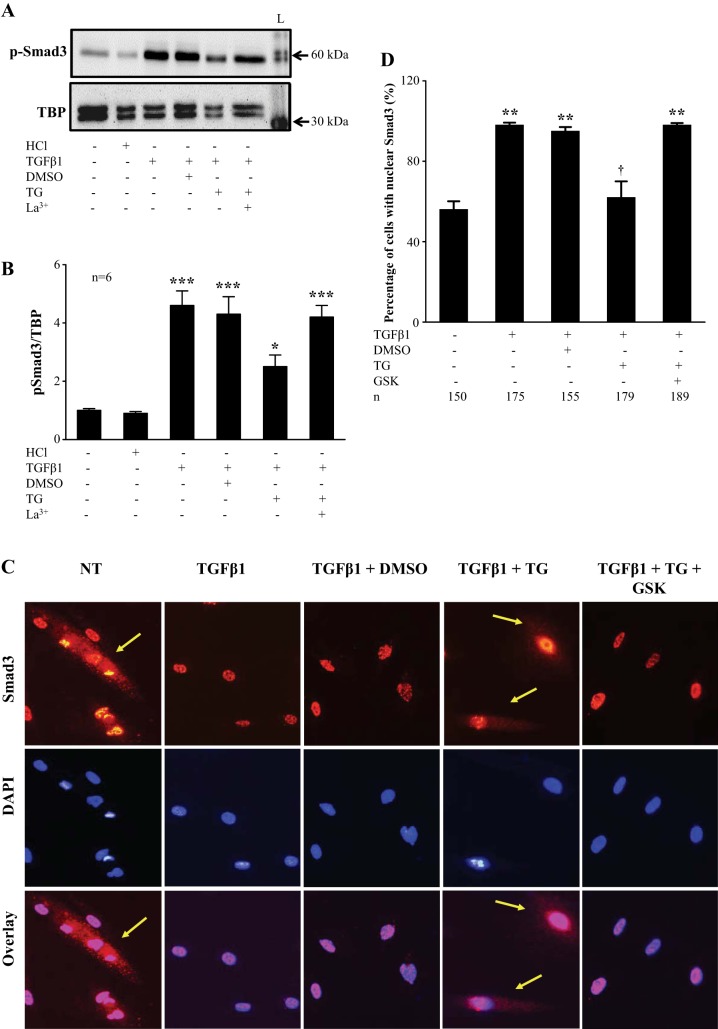
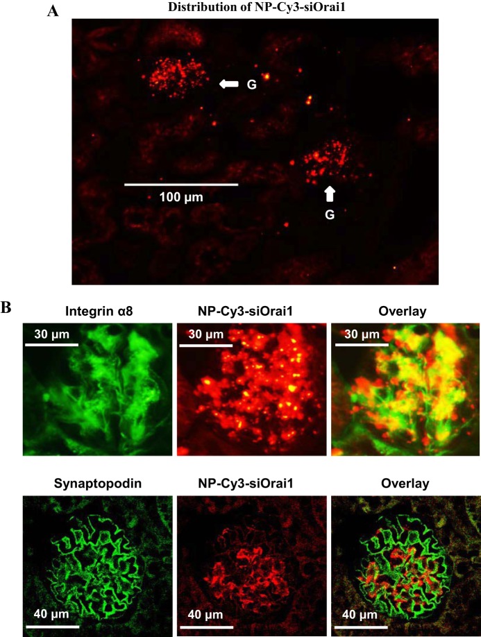
Our previous study demonstrated that the abundance of extracellular matrix proteins was suppressed by store-operated Ca2+ entry (SOCE) in mesangial cells (MCs). The present study was conducted to investigate the underlying mechanism focused on the transforming growth factor-β1 (TGF-β1)/Smad3 pathway, a critical pathway for ECM expansion in diabetic kidneys. We hypothesized that SOCE suppressed ECM protein expression by inhibiting this pathway in MCs. In cultured human MCs, we observed that TGF-β1 (5 ng/ml for 15 h) significantly increased Smad3 phosphorylation, as evaluated by immunoblot. However, this response was markedly inhibited by thapsigargin (1 µM), a classical activator of store-operated Ca2+ channels. Consistently, both immunocytochemistry and immunoblot showed that TGF-β1 significantly increased nuclear translocation of Smad3, which was prevented by pretreatment with thapsigargin. Importantly, the thapsigargin effect was reversed by lanthanum (La3+; 5 µM) and GSK-7975A (10 µM), both of which are selective blockers of store-operated Ca2+ channels. Furthermore, knockdown of Orai1, the pore-forming subunit of the store-operated Ca2+ channels, significantly augmented TGF-β1-induced Smad3 phosphorylation. Overexpression of Orai1 augmented the inhibitory effect of thapsigargin on TGF-β1-induced phosphorylation of Smad3. In agreement with the data from cultured MCs, in vivo knockdown of Orai1 specific to MCs using a targeted nanoparticle small interfering RNA delivery system resulted in a marked increase in abundance of phosphorylated Smad3 and in nuclear translocation of Smad3 in the glomerulus of mice. Taken together, our results indicate that SOCE in MCs negatively regulates the TGF-β1/Smad3 signaling pathway.

Keywords: Orai1; Smad3; mesangial cells; store-operated Ca2+ entry; transforming growth factor-β1.


Figures

Fig. 1.
Fig. 1.
Effect of store-operated Ca2+ entry (SOCE) on transforming growth factor-β1 (TGF-β1) secretion in cultured human mesangial cells (MCs). ELISA, showing TGF-β1 concentration in culture media. Confluent human MCs were incubated with serum-free DMEM for 72 h. One group was without any treatment (NT), and the other groups were treated with DMSO (1:1,000) or TG (1 μM) for 8 and 15 h before collection of media. DMSO and thapsigargin (TG) were present in the media throughout the period of treatment; n = no. of independent experiments.
Fig. 2.
Fig. 2.
SOCE inhibited TGF-β1-induced phosphorylation of Smad3 in cultured human MCs. A: representative Western blot, showing changes in abundance of phosphorylated Smad3 (p-Smad3) and total Smad3 (Smad3) proteins in different treatment groups. Human MCs were treated with recombinant human TGF-β1 (5 ng/ml) in the presence or absence of TG (1 μM) for 15 h. NT, cells without any treatment; HCl, 4 mM HCl with 0.1% BSA at 1:4,000, the vehicle control for TGF-β1; DMSO (1:1,000), the vehicle control for TG. α-Tubulin was used as the loading control. B: summary data showing changes in the ratio of p-Smad3 to Smad3 in different treatment groups. ***P < 0.001 vs. NT and HCl; **P < 0.01 vs. TGF-β1 and TGF-β1 + DMSO (n = no. of independent experiments).
Fig. 3.
Fig. 3.
Knockdown of Orai1 increased the TGF-β1-induced phosphorylation of Smad3. A: representative Western blot showing effect of knockdown of Orai1 on phosphorylated Smad3 (p-Smad3) and total Smad3 (Smad3) protein abundance. Human MCs were without transfection (UT) or transfected with scramble (Scr) or Orai1 small interfering (si)RNA (siOrai1). On day 3 after transfection cells were treated with TGF-β1 (5 ng/ml) in the presence or absence of TG (1 μM) for 15 h, α-tubulin was used as the loading control. L, protein ladder. B: summary data from experiments presented in A. The abundance of p-Smad3 is expressed as the ratio of p-Smad3 to Smad3. **P < 0.01 and ***P < 0.001 vs. UT; *P < 0.05 vs. TGF-β1, TGF-β1 + Scr, and TGF-β1 + siOrai1 + TG; #P < 0.05 vs. TGF-β1 + siOrai1 + TG; ##P < 0.01 vs. TGFβ1 + siOrai1 (n = no. of independent experiments).
Fig. 4.
Fig. 4.
Overexpression of Orai1 decreased the TGF-β1-induced phosphorylation of Smad3. A: representative Western blot showing phosphorylated Smad3 (p-Smad3) and total Smad3 (Smad3) protein abundance in human MCs in different groups. Human MCs were without transfection or transfected with yellow fluorescent protein (YFP) plasmid or mCherry-FLAG-Red Orai1 expression plasmid (Orai1). On day 2 after transfection, cells were treated with TGF-β1 (5 ng/ml) in the presence or absence of TG (1 μM) for 15 h. UT, cells without transfection or treatment; DMSO (1:1,000), vehicle control for TG. α-Tubulin was used as the loading control. B: summary data showing changes in p-Smad3/Smad3 ratio in different groups. ***P < 0.01 vs. UT; *P < 0.05 vs. TGF-β1, TGF-β1 + DMSO, and TGF-β1 + Orai1 + TG (n = no. of independent experiments). C: Western blot showing endogenous Orai1 and expressed Orai1 protein contents in human MCs transfected with YFP and Orai1. The expressed Orai1 protein was probed with primary antibody against Orai1 (top) and FLAG (middle). D: effect of overexpression of Orai1 on SOCE in human MCs. Fura-2 fluorescence ratiometry was used to assess the intracellular Ca2+ concentration ([Ca2+]i) in MCs without transfection (Untrans) or transfection with YFP plasmid or mCherry-FLAG-Red Orai1 expression plasmid (Orai1). SOCE was evaluated using a Ca2+ readdition protocol. TG (1 µM) was used to activate store-operated Ca2+ channels. **P < 0.01 compared with both Untrans and YFP groups; n = no. of cells analyzed in each group.
Fig. 5.
Fig. 5.
SOCE decreased TGF-β1-stimulated nuclear translocation of Smad3 in human MCs. A: representative Western blot showing phosphorylated Smad3 (p-Smad3) protein abundance in nuclear extracts of human MCs. Human MCs were either without treatment (NT) or treated with recombinant human TGF-β1 (5 ng/ml) in the presence or absence of TG (1 μM) or a selective blocker of SOCE, La3+ (5 μM), for 15 h. HCl, vehicle control for TGF-β1; DMSO, vehicle control for TG. TATA-binding protein (TBP) was used as the loading control for the nuclear proteins. L, protein ladder. B: summary data from experiments presented in A. ***P < 0.001 vs. NT and HCl; *P < 0.05 vs. TGF-β1, TGFβ1 + DMSO, and TGF-β1 + TG + La3+ (n = no. of independent experiments). C: representative images of immunofluorescence staining showing Smad3 expression in human MCs treated with TGF-β1 (5 ng/ml) in the presence or absence of TG (1 μM) or GSK-7975A (10 μM) for 15 h. NT, cells without treatment; DMSO, vehicle control for TG. Smad3 is shown in red. Nuclei were stained with 4′,6-diamidino-2-phenylindole and shown in blue. Purple indicates colocalization of Smad3 with nuclei. Arrows indicate distribution of Smad3 in the cytosol. D: summary data from 3 independent experiments showing %cells in which Smad3 was entirely localized in the nucleus in all cells counted. In each experiment, 3–4 fields were randomly selected and captured for analysis. **P < 0.01 compared with NT group; †P < 0.01 compared with groups of TGF-β1, TGF-β1 + DMSO, and TGF-β1 + TG + GSK. The numbers under each bar (n) represent the total nos. of cells analyzed from 5 image fields/experiment of 3 independent experiments.
Fig. 6.
Fig. 6.
Distribution of NP-Cy3-siOrai1 in MCs in mouse kidney. A: representative images from 3 mice showing localization of NP-Cy3-siOrai1 (red) in glomeruli (indicated by arrows) but not in tubules. Original magnification, ×200. B: localization of NP-Cy3-siOrai1 in MCs (top), but not in podocytes (bottom), represented from 3 mice. MCs and podocytes were stained with integrin-α8 (green) and synaptopodin (green), respectively. NP-Cy3-siOrai1 was shown as red signals. Original magnification, ×200.
Fig. 7.
Fig. 7.
Knockdown of Orai1 in MCs increased phosphorylation and nuclear translocation of Smad3 in mice. A: representative images for immunohistochemical staining of phosphorylated Smad3 (p-Smad3) on paraffin-embedded kidney sections from NP alone and NP-Cy3-siOrai1-injected mice. p-Smad3 staining is indicated in brown, whereas nuclei are shown in blue. Glomeruli are indicated by arrows. Original magnification, ×200. B: magnified images of the region indicated by dashed boxes in A. C: integrated density (ID) of p-Smad3 staining averaged from 3 NP-Con mice and 3 NP-Cy3-siOrai1-treated mice. The numbers in parentheses under each bar represent the no. of glomeruli counted from 5 sections/kidney. D: %cells without nuclear Smad3 in all cells counted in glomeruli, averaged from 3 NP-Con mice and 3 NP-Cy3-siOrai1-treated mice. The numbers in parentheses under each bar represent the no. of glomeruli counted from 5 sections/kidney. **P < 0.01, compared with NP-control.
Fig. 8.
Fig. 8.
The diagram illustrating the negative regulation of TGFβ1-Smad3 signaling by SOCE in MCs. p-Smad3: phosphorylated Smad3. Red line indicates inhibition and blue arrows indicate promotion of the pathway.

References

    1. Abboud HE. Mesangial cell biology. Exp Cell Res 318: 979–985, 2012. doi:10.1016/j.yexcr.2012.02.025. - DOI - PubMed
    1. Abdel-Wahab N, Wicks SJ, Mason RM, Chantry A. Decorin suppresses transforming growth factor-β-induced expression of plasminogen activator inhibitor-1 in human mesangial cells through a mechanism that involves Ca2+-dependent phosphorylation of Smad2 at serine-240. Biochem J 362: 643–649, 2002. doi:10.1042/bj3620643. - DOI - PMC - PubMed
    1. Abe H, Matsubara T, Iehara N, Nagai K, Takahashi T, Arai H, Kita T, Doi T. Type IV collagen is transcriptionally regulated by Smad1 under advanced glycation end product (AGE) stimulation. J Biol Chem 279: 14201–14206, 2004. doi:10.1074/jbc.M310427200. - DOI - PubMed
    1. Attisano L, Wrana JL. Smads as transcriptional co-modulators. Curr Opin Cell Biol 12: 235–243, 2000. doi:10.1016/S0955-0674(99)00081-2. - DOI - PubMed
    1. Bennett D, Alphey L. PP1 binds Sara and negatively regulates Dpp signaling in Drosophila melanogaster. Nat Genet 31: 419–423, 2002. doi:10.1038/ng938. - DOI - PubMed

Publication types

MeSH terms