Comparative Study
. 2017 Aug 11;292(32):13271-13283.
doi: 10.1074/jbc.M117.794578. Epub 2017 Jun 21.

An evolutionarily distinct family of polysaccharide lyases removes rhamnose capping of complex arabinogalactan proteins

Affiliations
Comparative Study

An evolutionarily distinct family of polysaccharide lyases removes rhamnose capping of complex arabinogalactan proteins

José Munoz-Munoz et al. J Biol Chem. .

Abstract

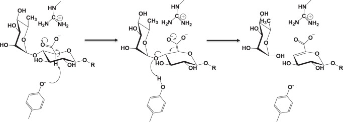
The human gut microbiota utilizes complex carbohydrates as major nutrients. The requirement for efficient glycan degrading systems exerts a major selection pressure on this microbial community. Thus, we propose that this microbial ecosystem represents a substantial resource for discovering novel carbohydrate active enzymes. To test this hypothesis we screened the potential enzymatic functions of hypothetical proteins encoded by genes of Bacteroides thetaiotaomicron that were up-regulated by arabinogalactan proteins or AGPs. Although AGPs are ubiquitous in plants, there is a paucity of information on their detailed structure, the function of these glycans in planta, and the mechanisms by which they are depolymerized in microbial ecosystems. Here we have discovered a new polysaccharide lyase family that is specific for the l-rhamnose-α1,4-d-glucuronic acid linkage that caps the side chains of complex AGPs. The reaction product generated by the lyase, Δ4,5-unsaturated uronic acid, is removed from AGP by a glycoside hydrolase located in family GH105, producing the final product 4-deoxy-β-l-threo-hex-4-enepyranosyl-uronic acid. The crystal structure of a member of the novel lyase family revealed a catalytic domain that displays an (α/α)6 barrel-fold. In the center of the barrel is a deep pocket, which, based on mutagenesis data and amino acid conservation, comprises the active site of the lyase. A tyrosine is the proposed catalytic base in the β-elimination reaction. This study illustrates how highly complex glycans can be used as a scaffold to discover new enzyme families within microbial ecosystems where carbohydrate metabolism is a major evolutionary driver.

Keywords: X-ray crystallography; carbohydrate processing; glycobiology; glycoside hydrolase; microbiome.


Conflict of interest statement

The authors declare that they have no conflicts of interest with the contents of this article

Figures

Figure 1.
Figure 1.
Biophysical and biochemical properties of BT0263 and BT3687. A, HPLC chromatogram showing GA only control (blue) and GA with the addition of BT0263 (green). The experimental conditions were 20 mm sodium phosphate buffer, 150 mm NaCl, pH 7.0; concentration of GA was 10 mm (refers to available rhamnose units) and enzyme was at 1 μm. B, pH-profile of BT0263. The experimental conditions were the same as in A for the substrate and enzyme concentrations. The buffers used were 20 mm sodium acetate, pH 4.0–5.0, 20 mm MES, pH 5.5–6.5, 20 mm sodium phosphate, pH 6.5–7.5, and 20 mm CAPS buffer, pH 7.5–9.0. The assays were monitored at a wavelength of UV A235 nm. C and D are HPLC chromatograms of GA only control (blue); GA treated with the GH43 exo-β1,3-d-galactanase BT0265 (red); GA treated with BT0265 and then BT0263 (green); GA treated sequentially with BT0265, BT0263, and the GH105 enzyme BT3687 (black). C is measured by pulsed amperometric detection, whereas D is measured via UV A235 nm. X indicates specific products, in addition to Rha, generated by BT0263 that are susceptible to BT3687, which specifically targets Δ4,5-UA generated by polysaccharide lyases. The experimental conditions were as in A.
Figure 2.
Figure 2.
Mass spectrometry of the oligosacchacaride. A major GA-derived oligosaccharide generated by the exo-β1,3-galatanase BT0265 (Fig. 1C) was purified by size exclusion chromatography. The oligosaccharide were untreated (A), incubated with BT0263 (B), or treated with BT0263 and then BT3687 (C). The experimental conditions for enzyme treatment were 20 mm sodium phosphate buffer, pH 7.0. The concentrations of substrate and enzyme were 1 mm and 1 μm, respectively.
Figure 3.
Figure 3.
The crystal structure of BACCELL_00875. A, schematic representation, ramped blue to red from the N to C terminus, of BACCELL_00875 showing the multidomain structure. B, active site of Baccell_00875 with residues implicated in substrate binding and catalysis shown in stick format. C, surface representation of the active site of Baccell_00875 in both conformations, loop closed (left) and loop open (right). In both cases, the residues implicated in stabilizing the conformation of this loop are shown as is the active site histidine whose position is dependent on the ionic lock. For reference the proposed catalytic tyrosine is also shown. D, surface representation of sequence conservation at the anterior and posterior sites of Baccell_00875. Residues were colored relative to their conservation within the PL27 family. Dark blue signifies amino acids that are invariant and cyan identifies residues that are 40–60% conserved. Red asterisk signifies the location of the active site.
Figure 4.
Figure 4.
The crystal structure of BT3687. A, schematic representation of BT3687 (beige) with the amino acids implicated in catalysis. The yellow amino acids, Asp116 and Asp160, are the proposed catalytic residues. The residues colored green and cyan are predicted to contribute to substrate binding. B, the conservation of residues in the −1 subsite (active site) between the GH105 enzymes BT3687 (green), NUGH105 (magenta), YTER (blue), YTER2 (beige), and the GH88 UGL (purple). The active site Trp56 of BT3687 (C) and the equivalent residue (Tyr41) in YTER (D) show how they are likely to interact with 4,5-UA-α1,2-l-Rha (left panel) and 4,5-UA-β-1,3-d-GalNAc (right panel), which were overlaid from structures with PDB codes 2GH4 and 2AHG, respectively.
Figure 5.
Figure 5.
Phylogenetic tree of the PL27 family. Four subgroups exhibit large evolutionary distances: Bacteroidetes (green subtree); Firmicutes and one Spirochaetes (orange subtree); Actinobacteria and a cohort pf Ascomycota (purple subtree); and Ascomycota-only (blue subtree). Labels on leaves indicate for each protein its species, its phylum, and the type of residue aligned to the catalytic Y. Split gene models that were manually fused for this analysis are indicated with the tag FUSED.
Figure 6.
Figure 6.
Proposed catalytic mechanism for PL27. The proposed catalytic mechanism, proceeding through an enolate transition state, with tyrosine acting as the catalytic acid/base and an arginine serving to stabilize the negative charge that is developed.

References

    1. Lozupone C. A., Stombaugh J. I., Gordon J. I., Jansson J. K., and Knight R. (2012) Diversity, stability and resilience of the human gut microbiota. Nature 489, 220–230 - PMC - PubMed
    1. Martens E. C., Lowe E. C., Chiang H., Pudlo N. A., Wu M., McNulty N. P., Abbott D. W., Henrissat B., Gilbert H. J., Bolam D. N., and Gordon J. I. (2011) Recognition and degradation of plant cell wall polysaccharides by two human gut symbionts. PLos Biol. 9, e1001221. - PMC - PubMed
    1. Cuskin F., Lowe E. C., Temple M. J., Zhu Y., Cameron E. A., Pudlo N. A., Porter N. T., Urs K., Thompson A. J., Cartmell A., Rogowski A., Hamilton B. S., Chen R., Tolbert T. J., Piens K., et al. (2015) Human gut Bacteroidetes can utilize yeast mannan through a selfish mechanism. Nature 517, 165–169 - PMC - PubMed
    1. Larsbrink J., Rogers T. E., Hemsworth G. R., McKee L. S., Tauzin A. S., Spadiut O., Klinter S., Pudlo N. A., Urs K., Koropatkin N. M., Creagh A. L., Haynes C. A., Kelly A. G., Cederholm S. N., Davies G. J., Martens E. C., and Brumer H. (2014) A discrete genetic locus confers xyloglucan metabolism in select human gut Bacteroidetes. Nature 506, 498–502 - PMC - PubMed
    1. Ndeh D., Rogowski A., Cartmell A., Luis A. S., Baslé A., Gray J., Venditto I., Briggs J., Zhang X., Labourel A., Terrapon N., Buffetto F., Nepogodiev S., Xiao Y., Field R. A., et al. (2017) Complex pectin metabolism by gut bacteria reveals novel catalytic functions. Nature 544, 65–70 - PMC - PubMed

Publication types

MeSH terms

LinkOut - more resources