

Comparison between Glioblastoma and Primary Central Nervous System Lymphoma Using MR Image-based Texture Analysis
- PMID: 28638001
- PMCID: PMC5760233
- DOI: 10.2463/mrms.mp.2017-0044
Comparison between Glioblastoma and Primary Central Nervous System Lymphoma Using MR Image-based Texture Analysis
Abstract
Purpose: To elucidate differences between glioblastoma (GBM) and primary central nervous system lymphoma (PCNSL) with MR image-based texture features.
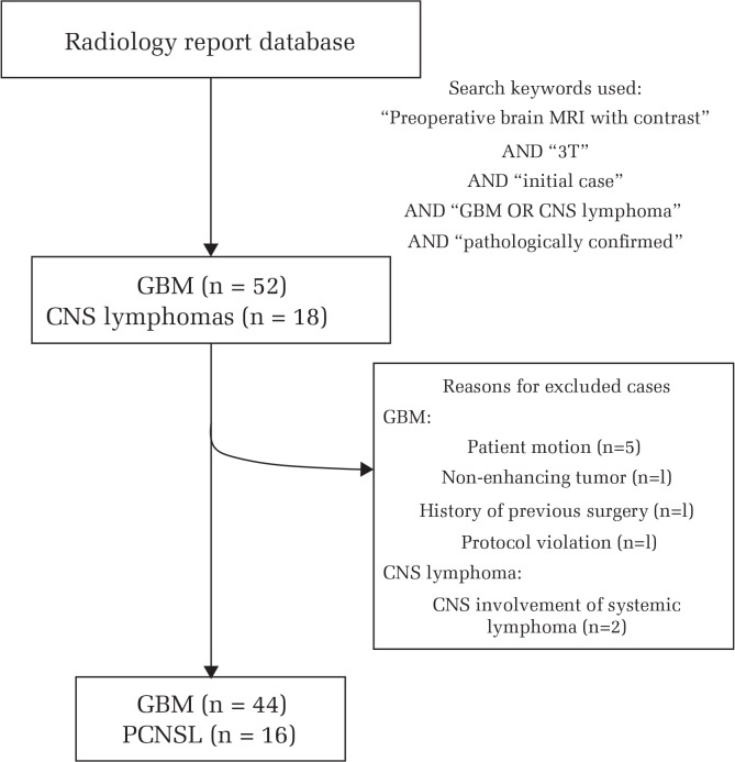
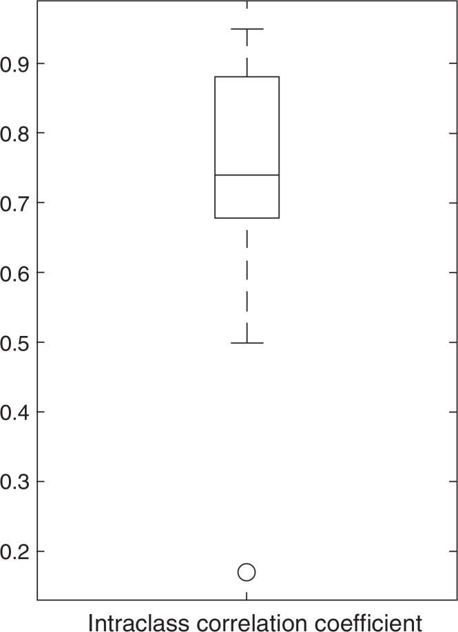
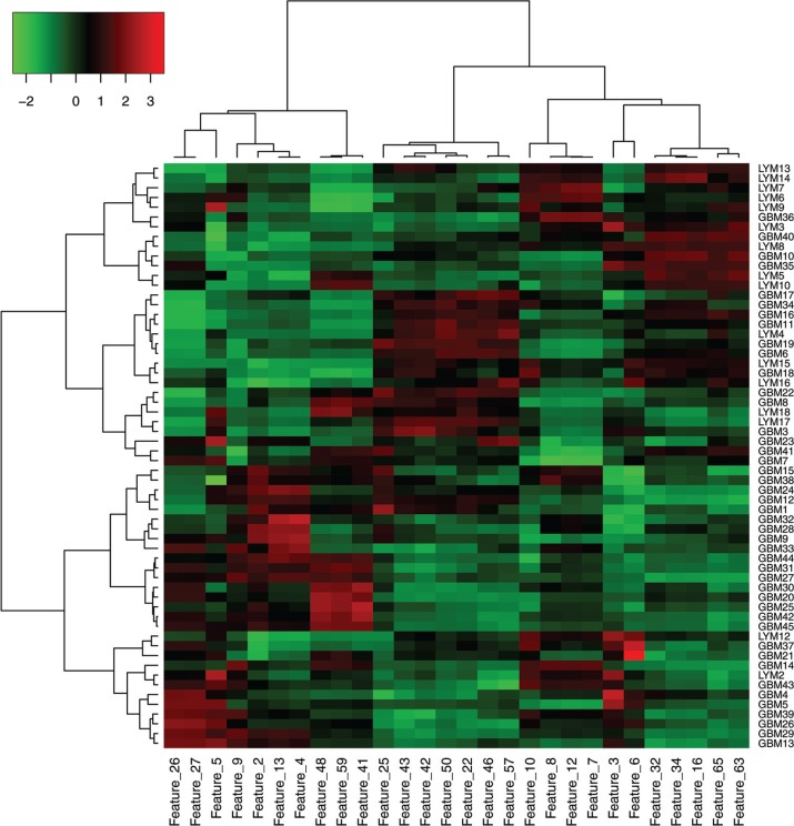
Methods: This was an Institutional Review Board (IRB)-approved retrospective study. Consecutive, pathologically proven, initially treated 44 patients with GBM and 16 patients with PCNSL were enrolled. We calculated a total of 67 image texture features on the largest contrast-enhancing lesion in each patient on post-contrast T1-weighted images. Texture analyses included first-order features (histogram) and second-order features calculated with gray level co-occurrence matrix, gray level run length matrix (GLRLM), gray level size zone matrix, and multiple gray level size zone matrix. All texture features were measured by two neuroradiologists independently and the intraclass correlation coefficients were calculated. Reproducible features with the intraclass correlation coefficients of greater than 0.7 were used for hierarchical clustering between the cases and the features along with unpaired t statistics-based comparisons under the control of false discovery rate (FDR) < 0.05. Principal component analysis (PCA) was performed to find the predominant features in evaluating the differences between GBM and PCNSL.
Results: Twenty-one out of the 67 features satisfied the acceptable intraclass correlation coefficient and the FDR constraints. PCA suggested first-order entropy, median, GLRLM-based run length non-uniformity, and run percentage as the distinguished features. Compared with PCNSL, run percentage and median were significantly lower, and entropy and run length non-uniformity were significantly higher in GBM.
Conclusions: Among MR image-based textures, first-order entropy, median, GLRLM-based run length non-uniformity, and run percentage are considered to enhance differences between GBM and PCNSL.
Keywords: glioblastoma; magnetic resonance imaging; primary central nervous system lymphoma; texture analysis.
Conflict of interest statement
The authors declare that they have no conflicts of interest.
Figures

References
-
- Louis DN, Ohgaki H, Wiestler OD, Cavenee WK. WHO classification of tumours of the central nervous system. Revised 4th ed International Agency for Research on Cancer (IARC); Lyon: 2016.
-
- Hochberg FH, Baehring JM, Hochberg EP. Primary CNS lymphoma. Nat Clin Pract Neurol 2007; 3:24–35. - PubMed
-
- Batchelor T, Loeffler JS. Primary CNS lymphoma. J Clin Oncol 2006; 24:1281–1288. - PubMed
-
- Stupp R, Mason WP, van den Bent MJ, et al. European organisation for research and treatment of cancer brain tumor and radiotherapy groups. national cancer institute of Canada clinical trials group Radiotherapy plus concomitant and adjuvant temozolomide for glioblastoma. N Engl J Med 2005; 352:987–996. - PubMed
-
- Kickingereder P, Wiestler B, Sahm F, et al. Primary central nervous system lymphoma and atypical glioblastoma: multiparametric differentiation by using diffusion-, perfusion-, and susceptibility-weighted MR imaging. Radiology 2014; 272:843–850. - PubMed
Publication types
MeSH terms
LinkOut - more resources
Full Text Sources
Other Literature Sources
Medical