Review
. 2017 Aug;18(8):459-469.
doi: 10.1038/nrn.2017.71. Epub 2017 Jun 22.

Neural circuits underlying thirst and fluid homeostasis

Affiliations
Review

Neural circuits underlying thirst and fluid homeostasis

Christopher A Zimmerman et al. Nat Rev Neurosci. 2017 Aug.

Abstract

Thirst motivates animals to find and consume water. More than 40 years ago, a set of interconnected brain structures known as the lamina terminalis was shown to govern thirst. However, owing to the anatomical complexity of these brain regions, the structure and dynamics of their underlying neural circuitry have remained obscure. Recently, the emergence of new tools for neural recording and manipulation has reinvigorated the study of this circuit and prompted re-examination of longstanding questions about the neural origins of thirst. Here, we review these advances, discuss what they teach us about the control of drinking behaviour and outline the key questions that remain unanswered.


Figures

Figure 1
Figure 1. Structure of the neural circuits underlying thirst and fluid homeostasis in mammals
a | The SFO is situated immediately dorsal to the third ventricle and contains intermingled populations of glutamatergic (SFOGLUT) and GABAergic (SFOGABA) neurons with opposing effects on drinking behavior. Optogenetic activation of SFOGLUT neurons (blue area, laser on, 30 min) stimulates intense drinking in hydrated mice, whereas optogenetic silencing of SFOGLUT neurons (green area, laser on, 15 min) suppresses drinking in dehydrated mice. In contrast, optogenetic activation of SFOGABA neurons (blue area, laser on, 15 min) suppresses drinking in dehydrated mice. b | SFO neurons innervate several brain regions involved in the regulation of fluid balance. SFOGLUT→MnPO/OVLT projections drive thirst, whereas the SFOGLUT→BNSTvl projection promotes sodium consumption. SFOGLUT→PVH/SON projections have not yet been functionally annotated with cell type-specificity, but classic models suggest that SFOGLUT→PVH/SON projections mediate secretion of AVP and OXT into the circulation and that the SFOGLUT→PVH projection may also modulate SNA and thereby alter blood pressure and heart rate. However, projections from MnPO/OVLT neurons to the PVH/SON also likely contribute to these neuroendocrine and autonomic responses. Projections from SFOGABA neurons to the MnPO/OVLT, as well as locally within the SFO, have not yet been functionally annotated. c | Sagittal illustration of the cell type-specific neural circuits underlying thirst and fluid homeostasis in the mouse brain. The LT consists of two circumventricular organs (SFO and OVLT) and an integratory structure (MnPO). Information about plasma osmolality, volume, and pressure enters the LT through specialized interoceptive neurons in the SFO and OVLT, some of which are intrinsically osmosensitive and AngII-sensitive (e.g., SFOGLUT neurons). The LT nuclei communicate with each other through an extensive network of bidirectional projections that has not yet been fully mapped with cell type-specificity. Outside the LT, SFOGLUT neurons project to the PVH, SON, and BNSTvl; projections from the MnPO and OVLT have not yet been mapped with cell type-specificity, however projections from these regions to the PVH and SON are well-established. SCNAVP neurons project to the OVLT and SON to mediate circadian regulation of thirst and AVP secretion, respectively. Information about plasma sodium enters the circuit through specialized aldosterone-sensitive NTSHSD2 neurons, which promote salt appetite and project to the pre-LC, PBN, and BNSTvl. Arch, archaerhodopsin; AVP, vasopressin; BNSTvl, ventrolateral part of the bed nucleus of the stria terminalis; ChR2, channelrhodopsin-2; MnPO, median preoptic nucleus; NTS, nucleus of the solitary tract; OVLT, organum vasculosum of the lamina terminalis; OXT, oxytocin; PBN, parabrachial nucleus; PP, posterior pituitary; pre-LC, pre-locus coeruleus; PVH, paraventricular hypothalamus; SCN, suprachiasmatic nucleus; SFO, subfornical organ; SNA, sympathetic nerve activity; SON, supraoptic nucleus.
Figure 2
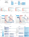
Figure 2. Anticipatory and homeostatic regulation of the thirst circuit
a | Challenges to fluid homeostasis, such as water deprivation, cause deviations in the composition of the blood. These deviations can include increases in plasma osmolality (Posmolality) and sodium (PNa), as well as decreases in plasma volume (Pvolume) and pressure that stimulate renin secretion by the kidneys and, consequently, AngII production (PAngII) in the blood. These homeostatic circulating signals are translated by the brain into counter-regulatory responses, including thirst, anorexia, vasopressin (PAVP) and oxytocin (POXT) secretion, and sympathetic nerve activation (SNA). While thirst is ultimately necessary to restore fluid homeostasis, this coordinated set of responses also promotes water retention (antidiuresis) and sodium excretion (natriuresis) by the kidneys, suppresses ingestion of food (and, therefore, sodium and other osmolytes), and modulates blood pressure and heart rate in order to maintain fluid homeostasis until water can be ingested. b | When thirsty animals are allowed to drink (red area), thirst and AVP secretion (blue lines) are rapidly inhibited before the composition of the blood (green lines) is corrected by ingested water. Historical experiments using esophageal/gastric fistulae and sham drinking suggest that this anticipatory regulation involves both immediate oropharyngeal and delayed visceral signals, however the neural mechanism underlying rapid anticipatory regulation of thirst and AVP secretion remained unexplored until recently. c | Fiber photometry recordings revealed that SFOGLUT neurons are rapidly inhibited during drinking to coordinate the anticipatory control of thirst and AVP secretion. In this example recording, a thirsty mouse (48 h water restriction) is given access to water. The inset highlights that rapid inhibition of SFOGLUT neurons is time-locked to individual drinking bouts (red areas). d | Fiber photometry recordings confirmed that SONAVP neurons are also rapidly inhibited during drinking. In this example recording, a thirsty mouse (24 h water restriction) is given access to water. The inset highlights that SONAVP neurons are transiently inhibited by water-predicting cues before drinking is initiated and that this inhibition is rapidly reset in the seconds immediately prior to water ingestion. The neural mechanism and physiological significance of this transient pre-ingestive inhibition remain unclear. e | In addition to homeostatic circulating signals (e.g., Posmolality, PNa, and PAngII) that were canonically thought to activate the LT (see Panel a), recent experiments have revealed that a diverse set of anticipatory oropharyngeal, visceral, and circadian signals also influence SFOGLUT neurons and the LT on different time-scales. This convergence of homeostatic and anticipatory signals allows SFOGLUT neurons and the LT to control a diverse set of behavioral, neuroendocrine, and autonomic outputs both in response to and in anticipation of deviations from fluid homeostasis. Data in Panel 2c is adapted from ref. 16. Data in Panel 2d is adapted from ref. 19.
Figure 3
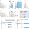
Figure 3. The thirst circuit monitors and controls feeding behavior
a | Eating potently stimulates prandial thirst, and most drinking therefore occurs in close temporal proximity to eating. Schematic is based on data in ref. 84. b | SFOGLUT neurons and SONAVP neurons are rapidly activated on both short (seconds) and long (minutes) time-scales during feeding to promote prandial thirst and AVP secretion. Schematics are based on data in refs. 16,. c | Dehydration potently suppresses food intake, and increases in plasma osmolality therefore inhibit feeding. Schematic is based on data in ref. 138. d | Dehydration anorexia causes hungry mice (24 h food restriction) to consume less food when water is absent (red line) than when water is available (black line). However, this suppression of food intake is completely alleviated by optogenetic silencing of SFOGLUT neurons during feeding (green line), indicating that activation of SFOGLUT neurons promotes dehydration anorexia in addition to prandial thirst. Schematic is based on data in ref. 16. e | Metabolic hormones secreted during feeding by the gastrointestinal tract-, pancreas-, and adipose tissue- have been proposed to regulate the electrical activity of SFO neurons ex vivo. However, it remains unclear whether such endocrine signals contribute to the natural coordination of eating and drinking by SFOGLUT neurons in vivo. f | Eating rapidly activates SFOGLUT neurons in order to promote prandial thirst and AVP secretion as well as dehydration anorexia. The mechanism of this activation remains unclear, but may involve learned or sensory neural signals or endocrine signals.

References

    1. Cannon WB. The physiological basis of thirst. Proceedings of the Royal Society B: Biological Sciences. 1918;90:283–301.
    1. Montgomery MF. The role of the salivary glands in the thirst mechanism. American Journal of Physiology. 1931;96:221–227.
    1. Leschke E. Ueber die durstempfindung. Archiv für Psychiatrie & Nervenkrankheiten. 1918;59:773–781.
    1. Gilman A. The relation between blood osmotic pressure, fluid distribution and voluntary water intake. American Journal of Physiology. 1937;120:323–328.
    1. Wolf AV. Osmometric analysis of thirst in man and dog. American Journal of Physiology. 1950;161:75–86. - PubMed

Publication types