. 2017 Jun 7:8:384.
doi: 10.3389/fphys.2017.00384. eCollection 2017.

Bile Acids Do Not Contribute to the Altered Calcium Homeostasis of Platelets from Rats with Biliary Cirrhosis

Affiliations

Bile Acids Do Not Contribute to the Altered Calcium Homeostasis of Platelets from Rats with Biliary Cirrhosis

Paola Romecín et al. Front Physiol. .

Abstract

Previously, we have found that intracellular calcium homeostasis is altered in platelets from an experimental model of liver cirrhosis, the bile-duct ligated (BDL) rat; these alterations are compatible with the existence of a hypercoagulable state and related to an enhanced intracellular calcium release evoked by thrombin and an increased amount of calcium stored in the intracellular organelles. In the present study we have investigated the role of bile acids in those alterations of the BDL cirrhotic model. Cholic acid (CA) or deoxycholic acid (DCA) did not change P-selectin expression or platelet aggregation in any group but elevated baseline platelet calcium levels. Incubation with both bile acids reduced calcium release after stimulation with thrombin in the absence of extracellular calcium. Pretreatment with CA but not with DCA reduced significantly thrombin-induced calcium entry in all three experimental groups. The capacitative calcium entry was also significantly lower in platelets pretreated with both bile acids. The simultaneous addition of thapsigargin and ionomycin to estimate the total amount of calcium in platelet internal stores was decreased by pretreatment with both CA and DCA, although these changes were significantly different in the control rats only with CA and in the BDL platelets with DCA. These results indicate that CA and DCA reduce calcium movements in platelets of control and BDL animals, thus suggesting that bile acids do not participate in the alterations observed in the BDL cirrotic model.

Keywords: bile-duct ligation; calcium signaling; capacitative calcium entry; cholestasis; fura-2; liver cirrhosis; thapsigargin; thrombin.


Figures

Figure 1
Figure 1
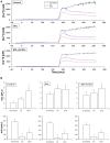
(A) Changes in [Ca2+]i in platelets of control (left column), BDL (middle column) and BDL with ascites rats (right column) in response to thrombin (0.3 U/ml, added at time 0). Top, in the absence of extracellular Ca2+. Bottom, in the presence of extracellular Ca2+. Data were obtained in untreated platelets and after incubation with CA or DCA (100 μM each). (B). Area under the curve of the calcium changes after thrombin administration (0.3 U/ml, added at time 0) in the absence (top panels) and in the presence (bottom panels) of extracellular Ca2+. Data were obtained in untreated platelets and after incubation with CA or DCA (100 μM each). Results are means ± S.E.M. *p < 0.05 vs. Untreated.
Figure 2
Figure 2
(A) Changes in [Ca2+]i after thapsigargin administration in the absence of extracellular Ca2+ (time 0 seconds) and after addition of calcium to allow for capacitative Ca2+ entry (time 180 s). Data were obtained in untreated platelets and after incubation with CA or DCA (100 μM). (B) Area under the curve of the calcium changes after thapsigargin administration in the absence of extracellular Ca2+ (top panels) and after addition of calcium to allow for capacitative Ca2+ entry (bottom panels). Data were obtained in untreated platelets and after incubation with CA or DCA (100 μM). Results are means ± S.E.M. *p < 0.05 vs. Untreated.
Figure 3
Figure 3
(A) Changes in [Ca2+]i after thapsigargin and ionomycin administration (added at time 0) to allow for estimation of total calcium in intracellular stores. Data were obtained in untreated platelets and after incubation with CA or DCA (100 μM). (B) Area under the curve of the calcium changes after thapsigargin and ionomycin to estimate total calcium in intracellular stores. Data were obtained in untreated platelets and after incubation with CA or DCA (100 μM). Data are means ± S.E.M. *p < 0.05 vs. Untreated.

References

    1. Annie-Jeyachristy S., Geetha A., Surendran R. (2008). Changes in the level of cytosolic calcium, nitric oxide and nitric oxide synthase activity during platelet aggregation: an in vitro study in platelets from normal subjects and those with cirrhosis. J. Biosci. 33, 45–53. 10.1007/s12038-008-0020-0 - DOI - PubMed
    1. Atucha N. M., Iyú D., Alcaraz A., Rosa V., Martínez-Prieto C., Ortiz M. C., et al. . (2007). Altered calcium signalling in platelets from bile-duct-ligated rats. Clin. Sci. 112, 167–174. 10.1042/CS20060226 - DOI - PubMed
    1. Atucha N. M., Iyu D., De Rycker M., Soler A., García-Estañ J. (2003). Altered calcium regulation in freshly isolated aortic smooth muscle cells from bile duct-ligated rats: Role of nitric oxide. Cell Calcium 33, 129–135. 10.1016/S0143-4160(02)00206-3 - DOI - PubMed
    1. Baele G., Beke R., Barbier F. (1980). In vitro inhibition of platelet aggregation by bile salts. Thromb. Haemost. 44, 62–64. - PubMed
    1. Bandi J. C., Poch E., García-Pagán J. C., Luca A., Jiménez W., Escorsell A., et al. . (1997). Platelet cytosolic calcium concentration in patients with liver cirrhosis. Relationship with hepatic and systemic hemodynamics. J. Hepatol. 27, 824–829. 10.1016/S0168-8278(97)80319-4 - DOI - PubMed

LinkOut - more resources