Review
. 2017 Jun 7:8:652.
doi: 10.3389/fimmu.2017.00652. eCollection 2017.

Expanding Role of T Cells in Human Autoimmune Diseases of the Central Nervous System

Affiliations
Review

Expanding Role of T Cells in Human Autoimmune Diseases of the Central Nervous System

Deepti Pilli et al. Front Immunol. .

Abstract

It is being increasingly recognized that a dysregulation of the immune system plays a vital role in neurological disorders and shapes the treatment of the disease. Aberrant T cell responses, in particular, are key in driving autoimmunity and have been traditionally associated with multiple sclerosis. Yet, it is evident that there are other neurological diseases in which autoreactive T cells have an active role in pathogenesis. In this review, we report on the recent progress in profiling and assessing the functionality of autoreactive T cells in central nervous system (CNS) autoimmune disorders that are currently postulated to be primarily T cell driven. We also explore the autoreactive T cell response in a recently emerging group of syndromes characterized by autoantibodies against neuronal cell-surface proteins. Common methodology implemented in T cell biology is further considered as it is an important determinant in their detection and characterization. An improved understanding of the contribution of autoreactive T cells expands our knowledge of the autoimmune response in CNS disorders and can offer novel methods of therapeutic intervention.

Keywords: T cell detection; autoantibodies; autoreactive T cells; central nervous system autoimmune diseases; multiple sclerosis; neuroimmunology.


Figures

Figure 1
Figure 1
Detection of human antigen-specific T cells with peptide-MHC (pMHC) multimer. Binding four pMHC monomers, for instance, via biotin–streptavidin interactions increases binding avidity between antigen and T cell receptor. This in turn enhances the sensitivity and specificity of antigen-specific T cells detected by flow cytometry analysis via the fluorescent streptavidin.
Figure 2
Figure 2
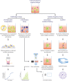
Functional assays commonly used in human T cell studies. Functional assays can be categorized into those that assess proliferative capacity of antigen-specific T cells and assays that analyze cytokine profiles upon T cell recognition of cognate antigen and subsequent activation. Proliferation assays can measure the amount of radioactive [3H]-thymidine incorporated into the DNA during cell division, with greater radioactivity indicating greater cell division. Alternatively, the level of fluorescence emitted by cells stained with carboxyfluorescein succinimidyl ester (CFSE) can be detected by flow cytometry, with greater number divisions correlating with lower fluorescence. In intracellular cytokine staining (ICS), protein secretion inhibitors, such as brefeldin A or monensin, allows for examination of cytokine production within a cell. Staining surface activation markers allows for phenotyping. Following fixation and permeabilization, the trapped intracellular cytokines are stained with fluorescent antibodies which can be detected via flow cytometry. Enzyme-linked immunospot (ELISPOT) is a popular method to assess cytokine secretion. The cytokine of interest secreted from an activated T cell is bound to a capture antibody on a PVDF bottom well. A biotinylated detection antibody also binds to the cytokine and facilitates the interaction between streptavidin-conjugated enzyme and its substrate to produce a color spot. Spots are quantified with a ELISPOT plate reader. Each spot represents one reactive cell.

References

    1. Goverman J. Autoimmune T cell responses in the central nervous system. Nat Rev Immunol (2009) 9(6):393–407.10.1038/nri2550 - DOI - PMC - PubMed
    1. Compston A, Coles A. Multiple sclerosis. Lancet (2008) 372(9648):1502–17.10.1016/S0140-6736(08)61620-7 - DOI - PubMed
    1. Hemmer B, Kerschensteiner M, Korn T. Role of the innate and adaptive immune responses in the course of multiple sclerosis. Lancet Neurol (2015) 14(4):406–19.10.1016/S1474-4422(14)70305-9 - DOI - PubMed
    1. Farrell MA, Droogan O, Secor DL, Poukens V, Quinn B, Vinters HV. Chronic encephalitis associated with epilepsy: immunohistochemical and ultrastructural studies. Acta Neuropathol (1995) 89(4):313–21.10.1007/BF00309624 - DOI - PubMed
    1. Dalmau J, Rosenfeld MR. Paraneoplastic syndromes of the CNS. Lancet Neurol (2008) 7(4):327–40.10.1016/S1474-4422(08)70060-7 - DOI - PMC - PubMed

LinkOut - more resources