Review
. 2017 Jun 7:4:37.
doi: 10.3389/fmolb.2017.00037. eCollection 2017.

Extracellular Vesicles in Renal Pathophysiology

Affiliations
Review

Extracellular Vesicles in Renal Pathophysiology

Margherita A C Pomatto et al. Front Mol Biosci. .

Abstract

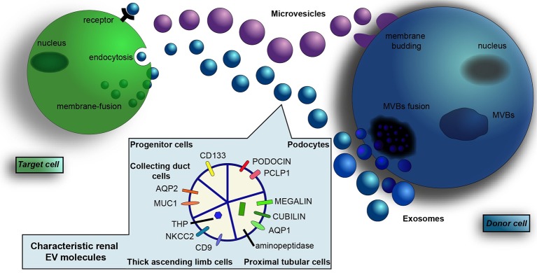
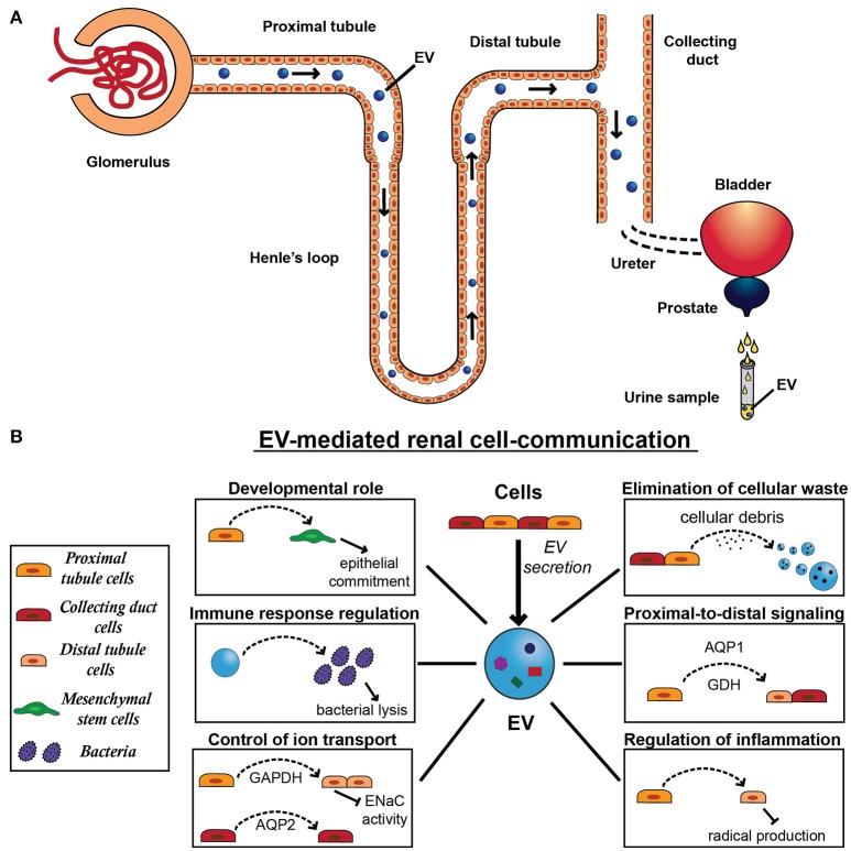
Extracellular vesicles are a heterogeneous population of microparticles released by virtually all living cells which have been recently widely investigated in different biological fields. They are typically composed of two primary types (exosomes and microvesicles) and are recently commanding increasing attention as mediators of cellular signaling. Indeed, these vesicles can affect recipient cells by carrying and delivering complex cargos of biomolecules (including proteins, lipids and nucleic acids), protected from enzymatic degradation in the environment. Their importance has been demonstrated in the pathophysiology of several organs, in particular in kidney, where different cell types secrete extracellular vesicles that mediate their communication with downstream urinary tract cells. Over the past few years, evidence has been shown that vesicles participate in kidney development and normal physiology. Moreover, EVs are widely demonstrated to be implicated in cellular signaling during renal regenerative and pathological processes. Although many EV mechanisms are still poorly understood, in particular in kidney, the discovery of their role could help to shed light on renal biological processes which are so far elusive. Lastly, extracellular vesicles secreted by renal cells gather in urine, thus becoming a great resource for disease or recovery markers and a promising non-invasive diagnostic instrument for renal disease. In the present review, we discuss the most recent findings on the role of extracellular vesicles in renal physiopathology and their potential implication in diagnosis and therapy.

Keywords: biomarkers; biomolecules; extracellular vesicles; intercellular communication; kidney; pathology; physiology.


Figures

Figure 1
Figure 1
Renal-derived extracellular vesicles. Extracellular vesicles (EVs) are a heterogeneous population of microparticles, mainly composed by exosomes and microvesicles. In particular, exosomes (in blue) are stored within multivesicular bodies (MVBs) of the late endosome and are released in the microenvironment after fusion with the cell membrane, whereas microvesicles (in violet) originate by direct budding from the cell surface. After their secretion, EVs exert their effects on adjacent or distant recipient cells in a pleiotropic manner, directly activating cell surface receptors, blending with cell membrane or by endocytic uptake and transferring their cargo inside cells. EVs contain a complex cargo of biomolecules that include proteins, surface receptors, lipids, transcription factors, genes, mRNAs, and miRNAs. Their content mirrors the cell of origin and EVs collected from urine contain proteins and transporters specific of renal and urogenital tract epithelial cells. In particular, the presence of podocin and podocalyxin (PCLP1) is characteristic of glomerular podocytes, whereas the expression of megalin, cubilin, aminopeptidase and aquaporin-1 (AQP1) indicate proximal tubular cell source. Moreover, EVs from the thick ascending limb of the Henle's loop contain Tamm Horsfall protein (THP), CD9, and type 2 Na-K-2Cl cotransporter (NKCC2). EVs from the collecting duct carry aquaporin-2 (AQP2) and mucin-1 (MUC1), whereas the expression of CD133 marker identify renal progenitor cells.
Figure 2
Figure 2
Extracellular vesicle secretion and physiological function in the kidney. (A) All cell types of the nephron that face the urinary space secrete EVs, starting from the glomerular podocytes through the proximal tubule, the limb of Henle, the distal tubule, and the collecting duct. After their secretion, EVs can be uptaken by downstream cells, influencing recipient cell behavior. Alternatively to their action on cells, EVs can cross the urinary tract and pass through following organs, including ureters, bladder, prostate, and urethra. EVs released by resident epithelial cells congregate with renal EVs and ultimately conveyed in the urine, providing a source of physiopathological markers of the urinary tract. (B) EV-mediated renal communication seems to be a physiological system of cell signaling and involves several EV roles, including elimination of cellular waste, proximal-to-distal signaling, developmental roles, control of ion transport, regulation of inflammation and immune response. In fact, EVs released by proximal tubule cells can be uptaken by distal tubule and collecting duct cells transferring tubular proteins, such as aquaporin-1 (AQP1) and the ammonium-generating enzyme glutaminase (GDH). EVs can also mediate the transfer of another aquaporin member, aquaporin-2 (AQP2) between cortical collecting duct cells. Moreover, by carrying active GAPDH, proximal tubule cells can regulate the renal transport of sodium through EVs, decreasing ENaC activity in distal tubule and collecting duct cells. Similarly, these EVs can also transfer anti-inflammatory message from proximal tubular cells exposed to dopamine receptor agonist and induce a decrease in cell radical production in distal tubular cells. Moreover, EVs derived from tubular cells are implicated in an important process for nephrogenesis and mediate the induction of the mesenchymal-to-epithelial transition (MET) in mesenchymal stem cells (MSCs). Finally, urinary EVs can induce bacterial lysis, contributing to the immune response in the urinary tract.

References

    1. Abdeen A., Sonoda H., El-Shawarby R., Takahashi S., Ikeda M. (2014). Urinary excretion pattern of exosomal aquaporin-2 in rats that received gentamicin. Am. J. Physiol. Ren. Physiol. 307, F1227–F12237. 10.1152/ajprenal.00140.2014 - DOI - PubMed
    1. Abdeen A., Sonoda H., Oshikawa S., Hoshino Y., Kondo H., Ikeda M. (2016). Acetazolamide enhances the release of urinary exosomal aquaporin-1. Nephrol. Dial. Transplant. 31, 1623–1632. 10.1093/ndt/gfw033 - DOI - PubMed
    1. Abid Hussein M. N., Nieuwland R., Hau C. M., Evers L. M., Meesters E. W., Sturk A. (2005). Cell-derived microparticles contain caspase 3 in vitro and in vivo. J. Thromb. Haemost. 3, 888–896. 10.1111/j.1538-7836.2005.01240.x - DOI - PubMed
    1. Alvarez M. L., Khosroheidari M., Ravi R. K., DiStefano J. K. (2012). Comparison of protein, microRNA, and mRNA yields using different methods of urinary exosome isolation for the discovery of kidney disease biomarkers. Kidney Int. 82, 1024–1032. 10.1038/ki.2012.256 - DOI - PubMed
    1. Alvarez S., Suazo C., Boltansky A., Ursu M., Carvajal D., Innocenti G., et al. . (2013). Urinary exosomes as a source of kidney dysfunction biomarker in renal transplantation. Transplant Proc. 45, 3719–3723. 10.1016/j.transproceed.2013.08.079 - DOI - PubMed