Review
. 2017:2017:5716835.
doi: 10.1155/2017/5716835. Epub 2017 May 30.

Abdominal Hernias, Giant Colon Diverticulum, GIST, Intestinal Pneumatosis, Colon Ischemia, Cold Intussusception, Gallstone Ileus, and Foreign Bodies: Our Experience and Literature Review of Incidental Gastrointestinal MDCT Findings

Affiliations
Review

Abdominal Hernias, Giant Colon Diverticulum, GIST, Intestinal Pneumatosis, Colon Ischemia, Cold Intussusception, Gallstone Ileus, and Foreign Bodies: Our Experience and Literature Review of Incidental Gastrointestinal MDCT Findings

G Di Grezia et al. Biomed Res Int. 2017.

Abstract

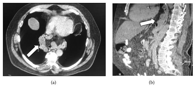
Incidental gastrointestinal findings are commonly detected on MDCT exams performed for various medical indications. This review describes the radiological MDCT spectrum of appearances already present in the past literature and in today's experience of several gastrointestinal acute conditions such as abdominal hernia, giant colon diverticulum, GIST, intestinal pneumatosis, colon ischemia, cold intussusception, gallstone ileus, and foreign bodies which can require medical and surgical intervention or clinical follow-up. The clinical presentation of this illness is frequently nonspecific: abdominal pain, distension, nausea, fever, rectal bleeding, vomiting, constipation, or a palpable mass, depending on the disease. A proper differential diagnosis is essential in the assessment of treatment and in this case MDCT exam plays a central rule. We wish that this article will familiarize the radiologist in the diagnosis of this kind of incidental MDCT findings for better orientation of the therapy.


Figures

Figure 1
Figure 1
(a) Axial plane, (b) coronal reconstruction, (c, d) sagittal reconstruction of abdominal MDCT exam showing a case of internal hernia, a left side paraduodenal hernia (white arrow).
Figure 2
Figure 2
(a, b) MDCT shows external left lumbar hernia (white arrow).
Figure 3
Figure 3
(a) Axial plane and (b) sagittal reconstruction of abdominal MDCT exam showing a case of mixed paradiaphragmatic hernia (white arrow).
Figure 4
Figure 4
(a) Coronal reconstruction, (b) axial plane, and (c) sagittal reconstruction of abdominal MDCT exam showing cases of gastric GIST (a, b), small bowel neoplasia (c) (white arrow), and postsurgical appearance of the lesions (d).
Figure 5
Figure 5
MDCT exam shows small bowel parietal pneumatosis (white arrow).
Figure 6
Figure 6
Colon ischemia axial images of MDCT showing bowel wall thickening corresponding to left colon (white arrow).
Figure 7
Figure 7
Cold intussusception axial images of MDCT showing the bowel pulled inward into itself (white arrow).
Figure 8
Figure 8
Axial images of MDCT show gallstone ileus (white arrow) in a typical location, the terminal ileum.
Figure 9
Figure 9
Axial images of MDCT (a) show a foreign body in a small bowel loop (white arrow), also detected in abdominal ultrasonography (b).

Similar articles

Cited by

References

    1. Waqas S., Johnson J.-O., Salastekar N., Maddu K. K., Khosa F. Incidental findings detected on abdomino-pelvic multidetector computed tomography performed in the acute setting. American Journal of Emergency Medicine. 2014;32(1):36–39. doi: 10.1016/j.ajem.2013.09.019. - DOI - PubMed
    1. Lassandro F., Iasiello F., Pizza N. L. Abdominal hernias: radiological features. World Journal of Gastrointestinal Endoscopy. 2011;3(6):110–117. doi: 10.4253/wjge.v3.i6.110. - DOI - PMC - PubMed
    1. Kohler A., Beldi G. Recurrence after hernia surgery: Complication or natural course? - PubMed
    1. Newsom B. D., Kukora J. S. Congenital and acquired internal hernias: unusual causes of small bowel obstruction. The American Journal of Surgery. 1986;152(3):279–285. doi: 10.1016/0002-9610(86)90258-8. - DOI - PubMed
    1. Takeyama N., Gokan T., Ohgiya Y., et al. CT of internal hernias. Radiographics. 2005;25(4):997–1015. doi: 10.1148/rg.254045035. - DOI - PubMed

MeSH terms

LinkOut - more resources