Review
. 2017 Sep;134(3):351-382.
doi: 10.1007/s00401-017-1739-1. Epub 2017 Jun 21.

Leukodystrophies: a proposed classification system based on pathological changes and pathogenetic mechanisms

Affiliations
Review

Leukodystrophies: a proposed classification system based on pathological changes and pathogenetic mechanisms

Marjo S van der Knaap et al. Acta Neuropathol. 2017 Sep.

Abstract

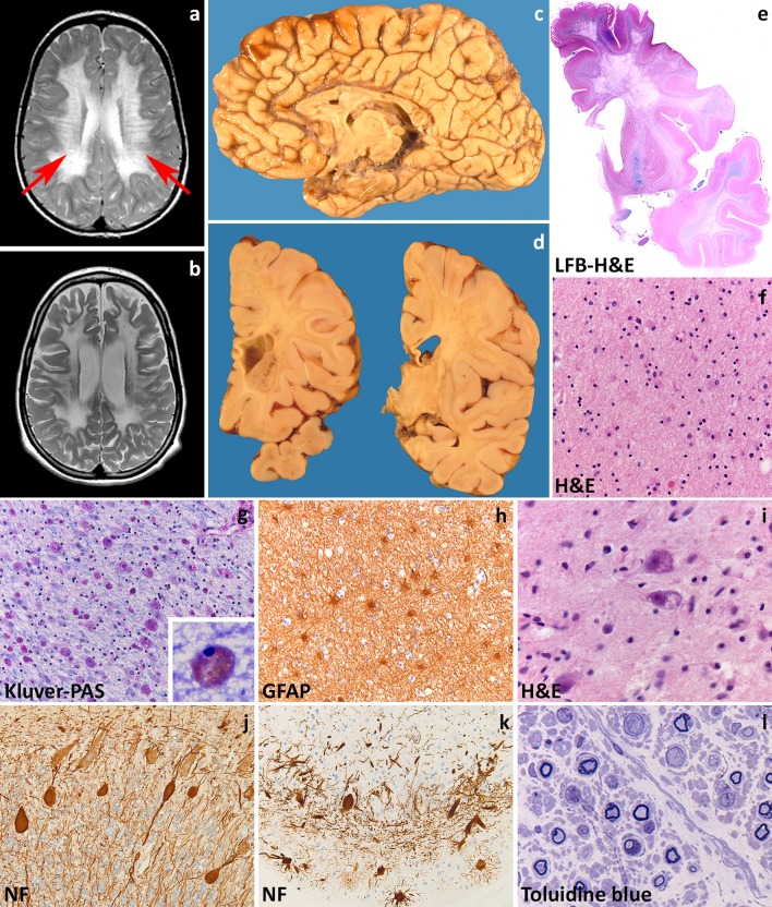
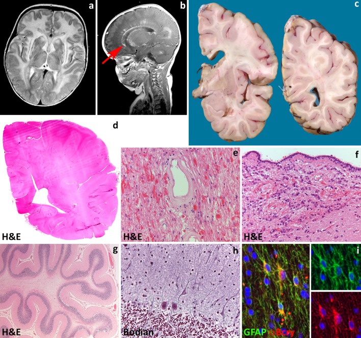
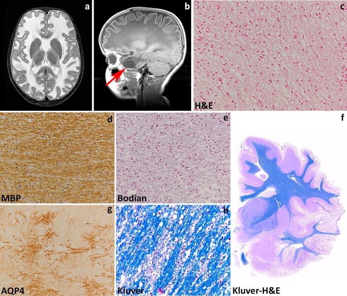
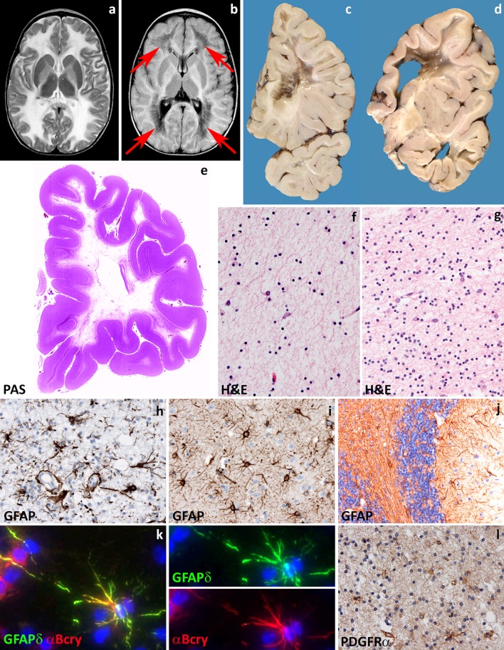
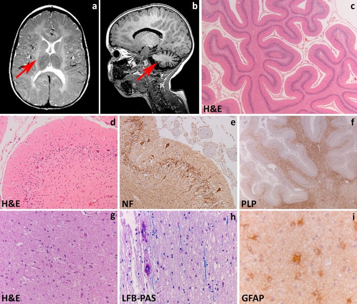
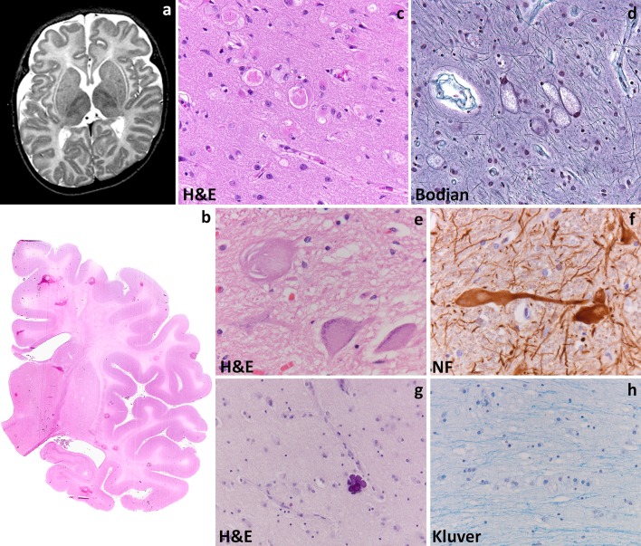
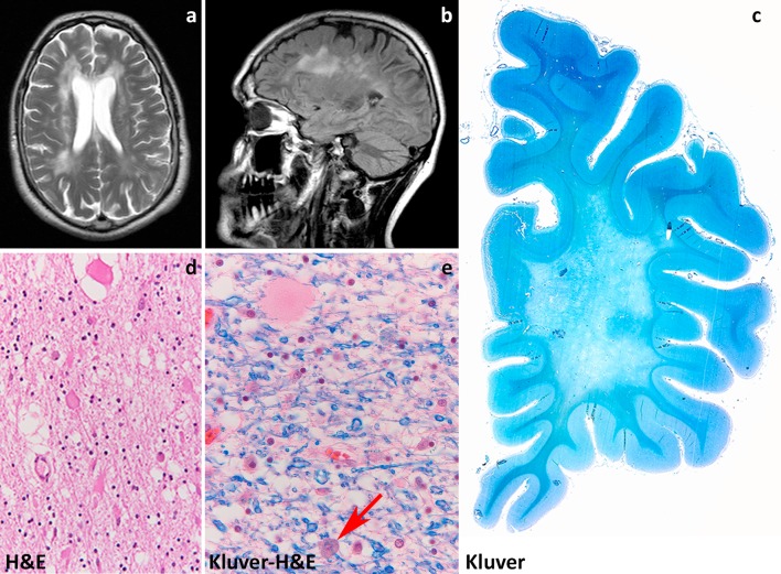
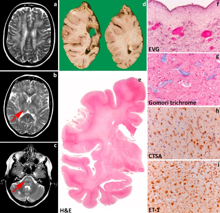
Leukodystrophies are genetically determined disorders characterized by the selective involvement of the central nervous system white matter. Onset may be at any age, from prenatal life to senescence. Many leukodystrophies are degenerative in nature, but some only impair white matter function. The clinical course is mostly progressive, but may also be static or even improving with time. Progressive leukodystrophies are often fatal, and no curative treatment is known. The last decade has witnessed a tremendous increase in the number of defined leukodystrophies also owing to a diagnostic approach combining magnetic resonance imaging pattern recognition and next generation sequencing. Knowledge on white matter physiology and pathology has also dramatically built up. This led to the recognition that only few leukodystrophies are due to mutations in myelin- or oligodendrocyte-specific genes, and many are rather caused by defects in other white matter structural components, including astrocytes, microglia, axons and blood vessels. We here propose a novel classification of leukodystrophies that takes into account the primary involvement of any white matter component. Categories in this classification are the myelin disorders due to a primary defect in oligodendrocytes or myelin (hypomyelinating and demyelinating leukodystrophies, leukodystrophies with myelin vacuolization); astrocytopathies; leuko-axonopathies; microgliopathies; and leuko-vasculopathies. Following this classification, we illustrate the neuropathology and disease mechanisms of some leukodystrophies taken as example for each category. Some leukodystrophies fall into more than one category. Given the complex molecular and cellular interplay underlying white matter pathology, recognition of the cellular pathology behind a disease becomes crucial in addressing possible treatment strategies.

Keywords: Astrocytes; Axons; Leukodystrophy; Microglia; Myelin; Oligodendrocytes.


Figures

Fig. 1
Fig. 1
Metachromatic leukodystrophy. a T2-weighted axial image of a 7-year-old child shows radiating stripes of tissue with preserved signal (arrows). The U-fibers are spared. b Follow-up T2-weighted axial image of the same child at 13 years shows a diffuse, bilateral and symmetric signal hyperintensity in the cerebral white matter. The U-fibers are no longer spared and the stripes are less well visible. There is a mild atrophy. c Sagittal cut of the brain of a 12-year-old child shows thinning of the corpus callosum and optic nerves. d On coronal sections through the brain of a 6-year-old child, the demyelinated white matter appears grayish and gelatinous. e Whole mount of a coronal section of a 10-year-old child stained with Luxol fast blue and Haematoxylin & Eosin shows diffuse loss of myelin in the frontal and temporal lobe with relative sparing of the U-fibers and internal capsule. f Haematoxylin & Eosin stain of the peripheral cerebral white matter shows tissue pallor, loss of oligodendrocytes and presence of foamy macrophages and reactive astrocytes. g Klüver-Periodic acid Schiff (PAS) stain of the same area shows loss of myelin and diffusely distributed macrophages filled with PAS-positive granular material. h Stain against the glial fibrillary acidic protein (GFAP) shows a moderate diffuse isomorphic astrogliosis. The inset shows a metachromatic macrophage stained with Toluidine blue. i Haematoxylin & Eosin stain of the thalamus shows accumulation of storage material in the cytoplasm of neurons. j, k Stain against neurofilaments (NF) shows axonal swellings (j) and dendritic varicosities (k) in the cerebellar cortical Purkinje cells. Note also the marked dropout of granular neurons in (k). l Toluidine blue stain of a semithin section of the sural nerve shows demyelination with accruing of foamy macrophages
Fig. 2
Fig. 2
Alexander disease. a T2-weighted axial image of a 9-month-old infant shows a diffuse, bilateral and symmetric signal hyperintensity with a clear frontal predominance. The abnormal white matter is also moderately swollen. Around the ventricles is a rim of lower signal intensity. The basal nuclei and thalami are abnormal in signal. b T1-weighted axial image of the same child shows contrast enhancement along the wall of the lateral ventricle and head of the caudate nucleus (arrow). c Coronal sections through the cerebral hemisphere of a 9-year-old child show that the white matter is intact, but slightly grayish. d Haematoxylin & Eosin stain of a whole mount shows loss of staining distinction between gray and white matter. e, f Haematoxylin & Eosin stain shows abundance of Rosenthal fibers around white matter blood vessels (e) and along the wall of the lateral ventricle (f). g Haematoxylin & Eosin stain of a cerebellar folium shows mild cortical atrophy and intense white matter pallor. h Bodian stain of the cerebellar cortex shows swelling of the Purkinje cell dendrites. i Double fluorescence stain reveals that glial fibrillary acidic protein (GFAP)-positive astrocytes also strongly express the heat shock protein α-B crystallin (α-Bcry)
Fig. 3
Fig. 3
Megalecephalic leukoencephalopathy with subcortical cysts. a T2-weighted axial image of an 8-year-old child with MLC1 mutations shows a diffuse, bilateral and symmetric signal hyperintensity in the cerebral white matter. The abnormal white matter is also mildly swollen. b T1-weighted sagittal image of the same child shows a subcortical cyst in the temporal pole (arrow). c Hematoxylin & Eosin stain of the subcortical white matter shows innumerable small vacuoles possibly crossed by thin tissue strands, indicative of intramyelinic oedema. d Stain against the major myelin protein myelin basic protein (MBP) shows normal amounts of myelin. e Bodian stain shows that axons are preserved. f Whole mount of a coronal section stained with Klüver-Haematoxylin & Eosin of a 30-year-old patient with a dominant GLIALCAM mutation shows complete integrity of the white matter. g In this patient, subcortical astrocytes strongly express the water channel Aquaporin 4 (AQP4), but the myelin amounts are normal and little intramyelinic oedema is present (h, Klüver stain)
Fig. 4
Fig. 4
Vanishing white matter. a T2-weighted axial image of a 1-year-old child shows a diffuse, bilateral and symmetric signal hyperintensity in the cerebral white matter extending to the internal and external capsules. b Fluid attenuated inversion recovery axial image of the same child shows loss of tissue in the periventricular and deep white matter (arrows). c, d Coronal cut of the brain of a 10- and a 6-year-old child confirms loss of the deeper cerebral white matter to a variable degree. The residual white matter appears grayish and gelatinous. There is a relative sparing of the internal capsule (d) and, in places, of the U-fibers. e Whole mount of a coronal section of a 10-year-old child stained with Haematoxylin & Eosin shows diffuse white matter rarefaction and cystic degeneration. f Haematoxylin & Eosin stain of the more affected frontal white matter shows marked tissue rarefaction with scarcity of astrocytes. g Haematoxylin & Eosin stain of the relatively spared cerebellar white matter shows some degree of tissue vacuolization and increased cellularity. h Stain against the glial fibrillary acidic protein (GFAP) of the more severely affected white matter shows dysmorphic astrocytes with short, blunt cell processes. i In the unaffected cerebral cortex, GFAP stain shows astrocytes with normal morphology. j In the cerebellar cortex, GFAP stain shows mislocalization of Bergmann glia to the molecular layer. k Double fluorescent stain shows that astrocytes robustly express the delta isoform of GFAP (GFAPδ) and the heat shock protein α-B crystallin (α-Bcry). l Stain against the oligodendrocyte precursor marker PDGFRα shows abundance of immunopositive cells in the relatively spared white matter
Fig. 5
Fig. 5
Hypomyelination with atrophy of the basal ganglia and cerebellum. a T2-weighted axial image of a 3-year-old child shows a mild, diffuse, bilateral and symmetric signal hyperintensity in the cerebral white matter. Note that the putamen has virtually disappeared (arrow). b T1-weighted sagittal image of the same child shows moderate cerebellar atrophy (arrow). The white matter is T1 hyperintense. The combination of mild T2 hyperintensity and T1 hyeprintensity is compatible with mild hypomyelination. c Haematoxylin & Eosin stain of a cerebellar folium shows cortical atrophy with thinning of the cortical granular layer. d Haematoxylin & Eosin stain of the cerebellar cortex reveals that loss of granular neurons may be severe and also accompanied by drop out of Purkinje cells. e Stain against neurofilaments (NF) shows swelling of cerebellar cortical Purkinje cells axons and dendrites. f Stain against the major myelin protein proteolipid protein (PLP) shows patchy lack of myelin in the cerebellar white matter. g Haematoxylin & Eosin stain of the caudate nucleus shows mild loss of neurons; the putamen could not be identified. h Luxol fast blue-Periodic acid Schiff (LFB-PAS) stain of the deeper white matter shows severe lack of myelin with decreased cellularity reflecting oligodendrocyte loss. i Stain against the glial fibrillary acidic protein (GFAP) of the white matter shows mild reactive gliosis with dividing astrocytes
Fig. 6
Fig. 6
GM1 gangliosidosis. a T2-weighted axial image of an 8-month-old infant shows a diffuse, bilateral and symmetric signal hyperintensity in the cerebral white matter extended to the U-fibers, but sparing the internal capsule. Note the mild signal abnormality of the basal nuclei and thalami. b Whole mount of a cerebral coronal section stained with Haematoxylin & Eosin shows diffuse cortical atrophy, and white matter pallor and atrophy with enlarged lateral ventricle and thinning of the corpus callosum. c Haematoxylin & Eosin stain of the frontal cortex shows accumulation of storage material in the cytoplasm of some neurons and reactive gliosis. d Bodian stain of the thalamus shows neuronal storage with ballooned cells also at this site. e Haematoxylin & Eosin stain of the anterior horn in the cervical spinal cord shows neuronal storage in the alpha motor neurons. f Stain against neurofilaments (NF) shows that the neuronal storage may be prominent at the level of the axon hillock, giving rise to meganeurites. g Haematoxylin & Eosin stain of the deep cerebral white matter shows lack of myelin and paucity of oligodendrocytes. h In the same area, a Klüver stain confirms the lack of myelin
Fig. 7
Fig. 7
Hereditary diffuse leukoencephalopathy with axonal spheroids. a T2-weighted axial image of a 50-year-old patient shows patchy, bilateral signal hyperintensities in the periventricular and deep cerebral white matter with posterior–frontal preponderance. b Patchy signal changes are also visible in a fluid attenuated inversion recovery sagittal image of the same patient. Note also the slight cerebral atrophy with widening of the sulci. c Whole mount of a cerebral coronal section stained with a Klüver shows confluent lack of myelin of the deep white matter with relative sparing of the U-fibers. d Haematoxylin & Eosin stain of the frontal white matter shows tissue rarefaction and axonal spheroids. e Klüver-Haematoxylin & Eosin stain of the parietal white matter shows lack of myelin, an axonal spheroid and a pigmented cell (arrow)
Fig. 8
Fig. 8
Cathepsin A-related arteriopathy with strokes and leukoencephalopathy. a T2-weighted axial image of a 52-year-old patient shows patchy and confluent signal hyperintensities in the periventricular and deep cerebral white matter. b, c T2-weighted axial image of the same patient show vascular lesions in the basal nuclei and thalami (b, arrow) and signal abnormalities in the basis of the pons (c, arrow). Two small infarctions in the right cerebellar hemisphere are visible (c). d Coronal sections of a cerebral hemisphere of 75-year-old patient show a cortico-subcortical infarct in the parietal lobe. The white matter appears relatively intact. e Whole mount of a coronal section of a 74-year-old patient stained with Haematoxylin & Eosin shows diffuse pallor of the periventricular and deep white matter with relative sparing of the subcortical areas and U-fibers. Note also small necrotic lesions in the periventricular white matter, basal nuclei and thalamus. f Elastic van Gieson stain of the periventricular white matter shows changes of the terminal arteriolar branches with asymmetric wall thickening and virtually complete lumen occlusion. g The same vascular changes are present in the deep white matter (Gomori trichrome). h Stain against cathepsin A (CTSA) shows strong immunoreactivity in white matter astrocytes. i In the same areas, astrocytes also robustly express endothelin-1 (ET-1)

References

    1. Abrams CK, Scherer SS. Gap junctions in inherited human disorders of the central nervous system. Biochem Biophys Acta. 2012;1818:2030–2047. doi: 10.1016/j.bbamem.2011.08.015. - DOI - PMC - PubMed
    1. Agamanolis DP. Pathology & genetics. Developmental neuropathology. City: ISN Neuropath; 2004.
    1. Alturkustani M, Keith J, Hazrati LN, Rademakers R, Ang LC. Pathologic staging of white matter lesions in adult-onset leukoencephalopathy/leukodystrophy with axonal spheroids. J Neuropathol Exp Neurol. 2015;74:233–240. doi: 10.1097/NEN.0000000000000168. - DOI - PubMed
    1. Anzil AP, Gessaga E. Late-life cavitating dystrophy of the cerebral and cerebellar white matter. A form of sudanophil leucodystrophy. Eur Neurol. 1972;7:79–94. doi: 10.1159/000114415. - DOI - PubMed
    1. Axelsson R, Roytta M, Sourander P, Akesson HO, Andersen O. Hereditary diffuse leucoencephalopathy with spheroids. Acta Psychiatr Scand Suppl. 1984;314:1–65. - PubMed

Publication types