. 2017 Nov;134(5):691-703.
doi: 10.1007/s00401-017-1743-5. Epub 2017 Jun 21.

Same-day genomic and epigenomic diagnosis of brain tumors using real-time nanopore sequencing

Affiliations

Same-day genomic and epigenomic diagnosis of brain tumors using real-time nanopore sequencing

Philipp Euskirchen et al. Acta Neuropathol. 2017 Nov.

Abstract

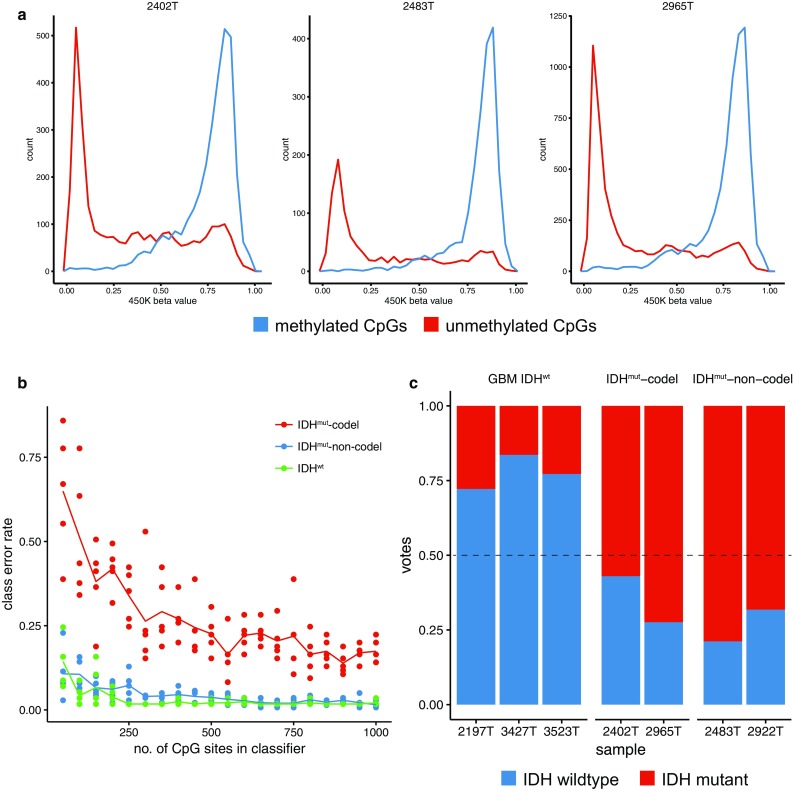
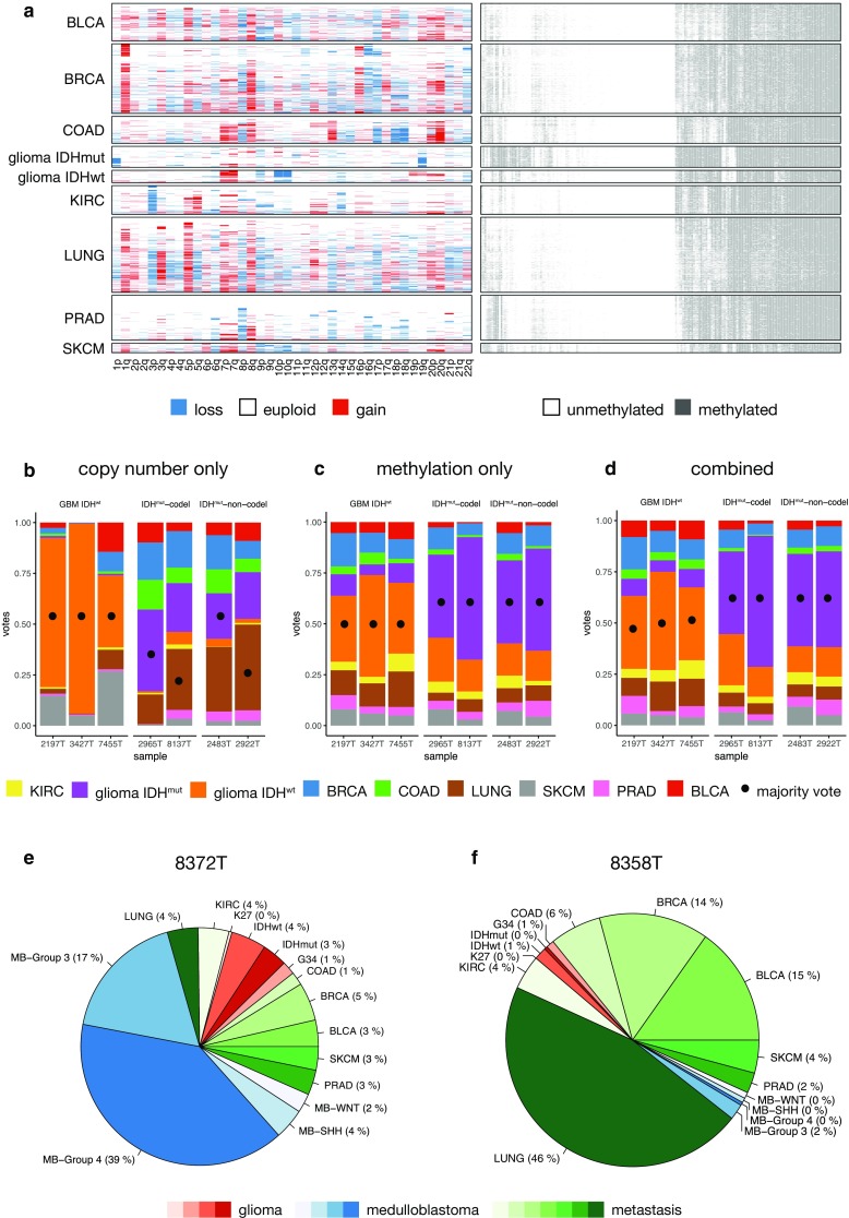
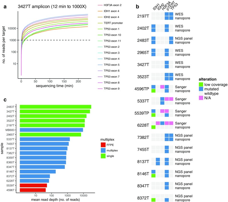
Molecular classification of cancer has entered clinical routine to inform diagnosis, prognosis, and treatment decisions. At the same time, new tumor entities have been identified that cannot be defined histologically. For central nervous system tumors, the current World Health Organization classification explicitly demands molecular testing, e.g., for 1p/19q-codeletion or IDH mutations, to make an integrated histomolecular diagnosis. However, a plethora of sophisticated technologies is currently needed to assess different genomic and epigenomic alterations and turnaround times are in the range of weeks, which makes standardized and widespread implementation difficult and hinders timely decision making. Here, we explored the potential of a pocket-size nanopore sequencing device for multimodal and rapid molecular diagnostics of cancer. Low-pass whole genome sequencing was used to simultaneously generate copy number (CN) and methylation profiles from native tumor DNA in the same sequencing run. Single nucleotide variants in IDH1, IDH2, TP53, H3F3A, and the TERT promoter region were identified using deep amplicon sequencing. Nanopore sequencing yielded ~0.1X genome coverage within 6 h and resulting CN and epigenetic profiles correlated well with matched microarray data. Diagnostically relevant alterations, such as 1p/19q codeletion, and focal amplifications could be recapitulated. Using ad hoc random forests, we could perform supervised pan-cancer classification to distinguish gliomas, medulloblastomas, and brain metastases of different primary sites. Single nucleotide variants in IDH1, IDH2, and H3F3A were identified using deep amplicon sequencing within minutes of sequencing. Detection of TP53 and TERT promoter mutations shows that sequencing of entire genes and GC-rich regions is feasible. Nanopore sequencing allows same-day detection of structural variants, point mutations, and methylation profiling using a single device with negligible capital cost. It outperforms hybridization-based and current sequencing technologies with respect to time to diagnosis and required laboratory equipment and expertise, aiming to make precision medicine possible for every cancer patient, even in resource-restricted settings.

Keywords: Brain tumor; Epigenomics; Glioma; Molecular neuropathology; Nanopore sequencing; Whole genome sequencing.


Conflict of interest statement

Funding

This work has been supported by Deutsche Forschungsgemeinschaft (EU 162/1-1 to PE), the program “Investissements d’avenir” (ANR-10-IAIHU-06 to AI), Institut Universitaire de Cancérologie (to AI), Ligue Nationale Contre le Cancer (to AI), Institut Carnot (to KL), and Fondation ARC pour la recherche sur le cancer (n°PJA 20151203562 to FB).

Conflict of interest

The authors declare no conflicts of interest.

Figures

Fig. 1
Fig. 1
Copy number profiling using nanopore low-pass whole genome sequencing. a Same-day workflows to simultaneously characterize copy number variation (CNV) and methylation profiles or single nucleotide variants, respectively. Tumor DNA is subjected to quality control (QC), and then, 250 ng input material is used for library preparation for either whole genome sequencing (WGS) or PCR-based deep amplicon sequencing. b Representative read length distribution of mapped reads. Note log scale on X axis. c Representative distribution of GC content of reads in comparison with the hg19 human reference genome. A randomly drawn subsample of the entire reference genome split into 1000 bp fragments is shown. d Copy number profile showing log2 transformed, normalized read counts per 1000 kbp window (grey) with running mean (red) and segmentation results (blue). e Comparison of nanopore WGS with matched SNP arrays. Heatmaps indicate copy number calls (losses and deletions in blue, and gains and amplifications in red) across the genome
Fig. 2
Fig. 2
Methylome profiling by nanopore sequencing of native tumor DNA. a Comparison of methylation calls from nanopore sequencing with matched Illumina 450K microarray-based data. Beta value distributions for CpG sites that were identified as unmethylated (red) or methylated (blue), respectively, by nanopore WGS are shown. b “Random taiga” simulation of classification error as a function of the number of randomly sampled CpG sites. Each dot represents the class-specific error rate of an ad hoc generated random forest using a random subset of N CpG sites (indicated on X axis) from the TCGA lower grade glioma Illumina 450K cohort as training set. Lines indicate the mean of five independent simulations. c Methylation profiles from nanopore sequencing discriminate IDH-mutant and wild-type tumors. Bar plots indicate vote distribution from ad hoc random forest classification. The TCGA low-grade glioma cohort was used as a training set. Illumina 450K-based beta values were dichotomized using >0.6 as threshold
Fig. 3
Fig. 3
Pan-cancer classification using copy number and methylation profiles. a Training set composed of TCGA samples from nine cancer entities using arm-level averaged copy number (CN) information (CN loss blue, CN gain red) and dichotomized methylation data. For illustration purposes, only 200 random CpG sites were sampled, clustered, and plotted. bd Classification of samples subjected to WGS using R9.4 flow cells using ad hoc random forests (500 trees per sample). Bar plots show vote distributions based on copy number only (b), methylation (c), or both modalities (d). e, f Methylation-based pan-cancer classification of medulloblastoma (e) and a brain metastasis of a lung adenocarcinoma (f). BRCA breast cancer, BLCA bladder urothelial carcinoma, COAD colon adenocarcinoma, KIRC kidney renal cell carcinoma, LUNG lung squamous cell and adenocarcinoma, SKCM skin cutaneous melanoma, PRAD prostate adenocarcinoma, MB medulloblastoma, K27 diffuse midline glioma H3 K27M mutant, G34 pediatric glioblastoma, H3 G34R mutant
Fig. 4
Fig. 4
Real-time amplicon sequencing of single nucleotide variants. a Representative coverage plot of target regions in IDH1, IDH2, H3F3A, TP53, and TERT promoter region over time. The time needed to achieve 1000X depth in all amplicons is indicated. Note log scale on Y axis. b Mean read depth over all amplicons in samples processed individually or as barcoded multiplex libraries. Of note, FFPE samples were sequenced as part of a multiplex library. c Comparison of selected variant calls from nanopore sequencing (filtered for coding or hotspot mutations with minimum allele frequency >0.2) with reference calls from Sanger or Illumina sequencing

References

    1. Abeshouse A, Ahn J, Akbani R, Ally A, Amin S, Andry CD, et al. The molecular taxonomy of primary prostate cancer. Cell. 2015;163:1011–1025. doi: 10.1016/j.cell.2015.10.025. - DOI - PMC - PubMed
    1. Akbani R, Akdemir KC, Aksoy BA, Albert M, Ally A, Amin SB, et al. Genomic classification of cutaneous melanoma. Cell. 2015;161:1681–1696. doi: 10.1016/j.cell.2015.05.044. - DOI - PMC - PubMed
    1. Breiman L. Random forests. Mach Learn. 2001;45:5–32. doi: 10.1023/A:1010933404324. - DOI
    1. Brennan CW, Verhaak RGW, McKenna A, Campos B, Noushmehr H, Salama SR, et al. The somatic genomic landscape of glioblastoma. Cell. 2013;155:462–477. doi: 10.1016/j.cell.2013.09.034. - DOI - PMC - PubMed
    1. Brat DJ, Verhaak RGW, Aldape KD, Yung WKA, Salama SR, Cancer Genome Atlas Research Network et al. Comprehensive, integrative genomic analysis of diffuse lower-grade gliomas. N Engl J Med. 2015;372:2481–2498. doi: 10.1056/NEJMoa1402121. - DOI - PMC - PubMed

Publication types