. 2017 Sep;134(3):459-473.
doi: 10.1007/s00401-017-1738-2. Epub 2017 Jun 21.

Parietal white matter lesions in Alzheimer's disease are associated with cortical neurodegenerative pathology, but not with small vessel disease

Affiliations

Parietal white matter lesions in Alzheimer's disease are associated with cortical neurodegenerative pathology, but not with small vessel disease

Kirsty E McAleese et al. Acta Neuropathol. 2017 Sep.

Abstract

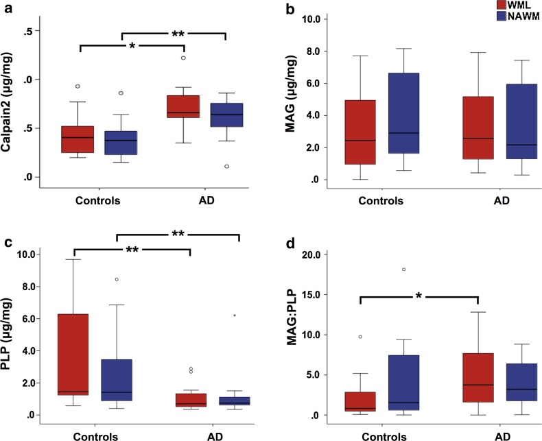
Cerebral white matter lesions (WML) encompass axonal loss and demyelination, and the pathogenesis is assumed to be small vessel disease (SVD)-related ischemia. However, WML may also result from the activation of Wallerian degeneration as a consequence of cortical Alzheimer's disease (AD) pathology, i.e. hyperphosphorylated tau (HPτ) and amyloid-beta (Aβ) deposition. WML seen in AD have a posterior predominance compared to non-demented individuals but it is unclear whether the pathological and molecular signatures of WML differ between these two groups. We investigated differences in the composition and aetiology of parietal WML from AD and non-demented controls. Parietal WML tissue from 55 human post-mortem brains (AD, n = 27; non-demented controls, n = 28) were quantitatively assessed for axonal loss and demyelination, as well as for cortical HPτ and Aβ burden and SVD. Biochemical assessment included Wallerian degeneration protease calpain and the myelin-associated glycoprotein (MAG) to proteolipid protein (PLP) ratio (MAG:PLP) as a measure of hypoperfusion. WML severity was associated with both axonal loss and demyelination in AD, but only with demyelination in controls. Calpain was significantly increased in WML tissue in AD, whereas MAG:PLP was significantly reduced in controls. Calpain levels were associated with increasing amounts of cortical AD-pathology but not SVD. We conclude that parietal WML seen in AD differ in their pathological composition and aetiology compared to WML seen in aged controls: WML seen in AD may be associated with Wallerian degeneration that is triggered by cortical AD-pathology, whereas WML in aged controls are due to ischaemia. Hence, parietal WML as seen on MRI should not invariably be interpreted as a surrogate biomarker for SVD as they may be indicative of cortical AD-pathology, and therefore, AD should also be considered as the main underlying cause for cognitive impairment in cases with parietal WML.

Keywords: Alzheimer’s disease; Hyperphosphorylated tau; Small vessel disease; Wallerian degeneration; White matter hyperintensity; White matter lesion.


Conflict of interest statement

Funding

The research was supported by the Alzheimer’s Society (Grant Number: AS-PG-2013-011). Tissue for this study was provided by the Newcastle Brain Tissue Resource, which is funded in part by a grant from the UK Medical Research Council (G0400074) and by Brains for Dementia research, a joint venture between Alzheimer’s Society and Alzheimer’s Research UK.

Conflict of interest

The authors declare that they have no conflict of interest.

Figures

Fig. 1
Fig. 1
a LFB stained sections were scanned and monochrome images were uploaded into Image-Pro Plus software program where the white matter was manually delineated to create a region of interest (green line). ai Using manual adjustment of the 8-bit grey scale threshold, total white matter area measured within the region of interest only. aii Using manual adjustment of the 8-bit grey scale threshold the WML was measured. Percentage area of WML per total white matter area was calculated and is expressed in the manuscript as WML area (WMLA). Scale bar 1 cm, valid for aaii
Fig. 2
Fig. 2
a WML were identified on LFB stained sections of parietal tissue and delineated from the NAWM by hand. This was used to identify the same WMLA on serial sections stained with Bielschowsky’s silver stain and SMI32. b A SMI32 stained section of parietal tissue indicating the five randomly selected areas for image capture in both the WMLA (black boxes) and NAWM (red boxes). bi 3 × 3 large image acquisition of SMI32-IR in WMLA. bii Magnified photomicroimage of SMI32 axonal positive pathology including discontinuous lines (white arrow) and end-bulbs axonal spheroid (black arrow). biii Image bi with applied bespoke SMI32 threshold including an example of artefact removal (white arrow). biv is image bii indicating discontinuous lines (white arrow) and end bull axonal spheroid (black arrow) with applied SMI32 threshold. Cellular nuclei were excluded from assessment (white arrow head). Mean area covered by IR was stated as a percentage of the total image area and the respective values are expressed as SMI32-IR. LFB luxol fast blue, Biel Bielschowsky’s silver stain, WMLA white matter lesion area, NAWM normal appearing white matter, IR immunoreactivity. Scale bar 50 mm, valid for all images in image a and image b; 100 μm, valid for bi, biii; 50 μm, valid for bii, biv
Fig. 3
Fig. 3
a AT8 stained parietal section indicating the four sample areas. ai 3 × 3 large image acquisition of cortex with AT8-IR. aii Image ai with applied bespoke threshold for AT8-IR only. Mean area covered by IR was stated as a percentage of the total image area and the respective values are expressed as AT8-IR. b H&E stained section of parietal tissue with deep white matter indicated (dashed line). Microphotoimages of up to eight individual white matter vessels were captured from the white matter for SI assessment. bi A microphotoimage of a white matter arteriole. SI was calculated using VasCalc software; [66] the internal and external diameters were measured three times to yield a SI value a mean SI value was calculated per section. IR immunoreactivity, SI sclerotic index, Dext external diameter, Dint internal diameter. Scale bar 1 cm, valid for images in a, b; 100 μm, valid for ai, aii; 50 μm, valid for bi
Fig. 4
Fig. 4
Scatter graphs show; ai negative correlations between WMLA and WML-BiA in AD cases only. aii Negative correlations between WMLA and NAWM-BiA in AD cases only. bi A positive correlation between WMLA and WML- and bii NAWM- LFB-IOD was seen in both AD and control cases. ci a negative correlation between WML-BiA and WML-LFB-IOD in AD cases only. cii A negative correlation between NAWM-BiA and NAWM-LFB-IOD was seen in AD cases only. p values and associated correlation coefficients are shown in main text. WML white matter lesion, NAWM normal appearing white matter, WMLA white matter lesion area, BiA Bielschowsky’s area, LFB-IOD luxol fast blue integrated optical density
Fig. 5
Fig. 5
Box plots show; a Calpain2 was significantly higher in the WML tissue of AD cases compared to controls. b No significant difference was seen in WML-MAG between AD and control cases. c PLP was significantly lower in the WML tissue of AD cases compared to controls. d MAG:PLP was significantly lower in the WML tissue of control compared to AD cases. *p < 0.05; ***p < 0.001. Exact p values are shown in main text. AD Alzheimer’s disease, MAG myelin associated glycoprotein, PLP proteolipid protein

References

    1. Acosta-Cabronero J, Williams GB, Pengas G, Nestor PJ. Absolute diffusivities define the landscape of white matter degeneration in Alzheimer’s disease. Brain. 2010;133:529–539. doi: 10.1093/brain/awp257. - DOI - PubMed
    1. Agosta F, Pievani M, Sala S, Geroldi C, Galluzzi S, Frisoni GB, et al. White matter damage in Alzheimer disease and its relationship to gray matter atrophy. Radiology. 2011;258:853–863. doi: 10.1148/radiol.10101284. - DOI - PubMed
    1. Amlien IK, Fjell AM, Walhovd KB, Selnes P, Stenset V, Grambaite R, et al. Mild cognitive impairment: cerebrospinal fluid tau biomarker pathologic levels and longitudinal changes in white matter integrity. Radiology. 2013;266:295–303. doi: 10.1148/radiol.12120319. - DOI - PubMed
    1. Attems J, Jellinger KA. Only cerebral capillary amyloid angiopathy correlates with Alzheimer pathology—a pilot study. Acta Neuropathol. 2004;107:83–90. doi: 10.1007/s00401-003-0796-9. - DOI - PubMed
    1. Barber R, Scheltens P, Gholkar A, Ballard C, McKeith I, Ince P, et al. White matter lesions on magnetic resonance imaging in dementia with Lewy bodies, Alzheimer’s disease, vascular dementia, and normal aging. J Neurol Neurosurg Psychiatry. 1999;67:66–72. doi: 10.1136/jnnp.67.1.66. - DOI - PMC - PubMed