. 2018 Jan;91(1):5-16.
doi: 10.1111/cbdd.13053. Epub 2017 Jul 19.

Mapping the allosteric sites of the A2A adenosine receptor

Affiliations

Mapping the allosteric sites of the A2A adenosine receptor

Alisha D Caliman et al. Chem Biol Drug Des. 2018 Jan.

Abstract

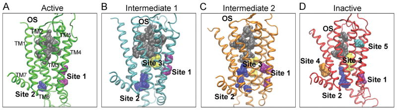
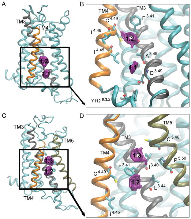
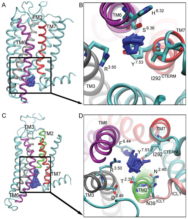
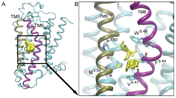
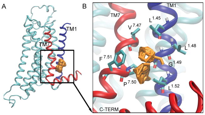
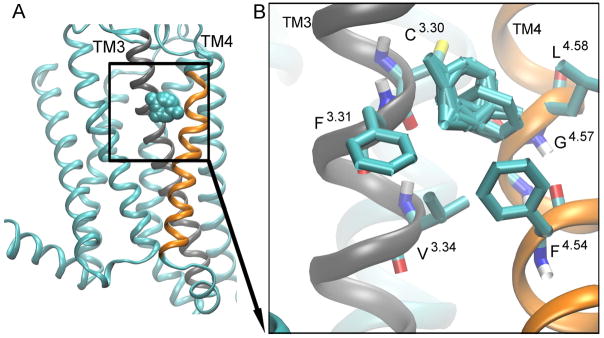
The A2A adenosine receptor (A2A AR) is a G protein-coupled receptor that is pharmacologically targeted for the treatment of inflammation, sepsis, cancer, neurodegeneration, and Parkinson's disease. Recently, we applied long-timescale molecular dynamics simulations on two ligand-free receptor conformations, starting from the agonist-bound (PDB ID: 3QAK) and antagonist-bound (PDB ID: 3EML) X-ray structures. This analysis revealed four distinct conformers of the A2A AR: the active, intermediate 1, intermediate 2, and inactive. In this study, we apply the fragment-based mapping algorithm, FTMap, on these receptor conformations to uncover five non-orthosteric sites on the A2A AR. Two sites that are identified in the active conformation are located in the intracellular region of the transmembrane helices (TM) 3/TM4 and the G protein-binding site in the intracellular region between TM2/TM3/TM6/TM7. Three sites are identified in the intermediate 1 and intermediate 2 conformations, annexing a site in the lipid interface of TM5/TM6. Five sites are identified in the inactive conformation, comprising a site in the intracellular region of TM1/TM7 and in the extracellular region of TM3/TM4 of the A2A AR. We postulate that these sites on the A2A AR be screened for allosteric modulators for the treatment of inflammatory and neurological diseases.

Keywords: GPCR; A2A adenosine receptor; FTMap; G protein-coupled receptors; allostery; fragment mapping.


Conflict of interest statement

Conflict of Interest

The authors declare that there are no conflicts of interests.

Figures

Figure 1
Figure 1. Probe Occupancy Per Residue during the Molecular Dynamic Simulations
Probe occupancies for the agonist-bound apo simulation [A], and the antagonist-bound apo simulation [B] are shown. Binding site residues are shown in red, non-orthosteric residues are shown in blue, and black boxes represent the transmembrane regions. ICL1 is located in between TM1 and TM2. ECL1 is located between TM2 and TM3. ICL2 is located between TM3 and TM4. ECL2 is located between TM4 and TM5. ICL3 is located between TM5 and TM6, and ECL3 is located between TM6 and TM7.
Figure 2
Figure 2. The Hot Spots on the A2AAR
The hot spots for probe-binding in the active (green) [A], intermediate 1 (cyan) [B], intermediate 2 (orange) [C], and inactive (red) [D] conformers are shown. The orthosteric site (OS) is shown in gray, site 1 in purple, site 2 in blue, site 3 yellow, site 4 in orange, and site 5 in cyan.
Figure 3
Figure 3. Hot spot 1: ‘The Intracellular Crevice’
is located in the intracellular region between TM3 (gray), TM4 (orange) and TM5 (tan). The probes in the active [A] and the intermediate 1 conformers [C] are shown as purple spheres, with the numbers ‘1.1’, ‘1.2’ and ‘1.3’ denoting different regions of the pocket. The key interacting residues for the active clusters [B] and intermediate 1 conformers [D] are shown as bonds, and the probes that bind to this site are shown as purple bonds.
Figure 4
Figure 4. Hot spot 2: ‘The G Protein-Coupling Site’
is located between TM2 (green), TM3 (gray), TM6 (purple), and TM7 (red). The probes in the active [A] and inactive [C] conformers are shown as blue spheres. The key interacting residues for active conformers [B] and inactive conformers [D] are shown as bonds, and the probes that bind to this site are shown as blue bonds.
Figure 5
Figure 5. Hot spot 3: ‘The Lipid interface’
is located between TM5 (tan) and TM6 (purple). The probes are shown as yellow spheres [A]. The key interacting residues are shown as bonds [B], and the probes that bind to this site are shown as yellow bonds.
Figure 6
Figure 6. Hot spot 4: ‘The C-Terminus Cleft’
is located between TM1 (blue), TM7 (red), and TM8 (red). The probes are shown as orange spheres [A]. The key interacting residues are shown as bonds [B], and the probes that bind to this site are shown as orange bonds.
Figure 7
Figure 7. Hot spot 5: ‘The Extracellular Cleft’
is located between TM3 (gray), and TM4 (orange) helices. The probes are shown as cyan spheres [A]. The key interacting residues are shown as bonds [B], and the probes that bind to this site are shown as cyan bonds.

References

    1. Thiel M, Caldwell CC, Sitkovsky MV. The critical role of adenosine A2A receptors in downregulation of inflammation and immunity in the pathogenesis of infectious diseases. Microbes and Infection. 2003;5(6):515–526. - PubMed
    1. Sivak KV, et al. Adenosine A2A receptor as a drug target for treatment of sepsis. Molecular Biology. 2016;50(2):200–212. - PubMed
    1. Cekic C, Linden J. Purinergic regulation of the immune system. Nature Reviews Immunology. 2016;16(3):177–192. - PubMed
    1. Chen JF, Eltzschig HK, Fredholm BB. Adenosine receptors as drug targets — what are the challenges? Nature Reviews Drug Discovery. 2013;12(4):265–286. - PMC - PubMed
    1. Pinna A. Adenosine A2A receptor antagonists in Parkinson’s disease: progress in clinical trials from the newly approved istradefylline to drugs in early development and those already discontinued. CNS Drugs. 2014;28(5):455–474. - PubMed

Publication types

MeSH terms

Substances