. 2017 Jun 22:8:15877.
doi: 10.1038/ncomms15877.

MicroRNA-34a dependent regulation of AXL controls the activation of dendritic cells in inflammatory arthritis

Affiliations

MicroRNA-34a dependent regulation of AXL controls the activation of dendritic cells in inflammatory arthritis

Mariola Kurowska-Stolarska et al. Nat Commun. .

Abstract

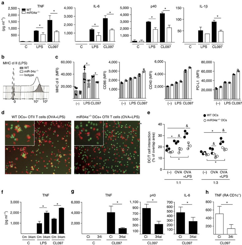
Current treatments for rheumatoid arthritis (RA) do not reverse underlying aberrant immune function. A genetic predisposition to RA, such as HLA-DR4 positivity, indicates that dendritic cells (DC) are of crucial importance to pathogenesis by activating auto-reactive lymphocytes. Here we show that microRNA-34a provides homoeostatic control of CD1c+ DC activation via regulation of tyrosine kinase receptor AXL, an important inhibitory DC auto-regulator. This pathway is aberrant in CD1c+ DCs from patients with RA, with upregulation of miR-34a and lower levels of AXL compared to DC from healthy donors. Production of pro-inflammatory cytokines is reduced by ex vivo gene-silencing of miR-34a. miR-34a-deficient mice are resistant to collagen-induced arthritis and interaction of DCs and T cells from these mice are reduced and do not support the development of Th17 cells in vivo. Our findings therefore show that miR-34a is an epigenetic regulator of DC function that may contribute to RA.


Conflict of interest statement

The authors declare no competing financial interests.

Figures

Figure 1
Figure 1. MiR-34a expression is upregulated in DCs from patients with RA.
(a) CD1c+ DCs were FACS-sorted from PB of healthy donors (HD, n=6), and from PB (n=6) and SF (n=6) of RA patients with established RA of >2 years duration. Compared to HD, miR-34a expression was upregulated in PB CD1c+ in RA patients (*P<0.05, Mann–Whitney U-test); and further upregulated in matched SF (**P<0.01, paired t-test). There was no significant difference (ns) in miR-34c expression between these groups. (b) miR-34a expression was upregulated in PB CD1c+ cells in patients (n=14) with early RA <6 months from diagnosis (eRA), as compared to age-matched HD (n=6) (*P<0.05, Kolmogorov–Smirnov test). (c) MiR-34a expression was upregulated in CD1c+ DCs FACS sorted from dispersed RA synovial membrane (SM, n=9) as compared to RA PB (n=19) and HD PB (n=10) cells (**P<0.01 Kruskal–Wallis test and Dunn’s multiple comparison test). Data presented as median and inter-quartile range boxplots; and for c, each dot represents single sample from naive (naive-to-treatment); MTX resistant (resistant to methotrexate treatment); TNFi resistant (resistant to combined therapy of methotrexate and TNF inhibitors). Demographic and clinical information for samples in ab is presented in Supplementary Table 1 (Cohort 1); and for samples in c is presented in Supplementary Table 2 (Cohort 2). ns, nonsignificant.
Figure 2
Figure 2. MiR-34a expression is regulated by GM-CSF and TLR ligands.
(a) A representative FACS-gating strategy for sorting healthy donor human blood monocyte subsets. (b) Each monocyte subset expressed low copy numbers of miR-34a relative to let-7a housekeeping control (c), which increased following differentiation with GM-CSF (50 ng ml−1) for 3 and 7 days. Data in b are presented as a dot plot with dotted lines joining the cells from the same donor for clarity. (d) Monocyte-derived DCs (n=3) were stimulated with TLR agonists CL097 (TLR7/8; 1 μg ml−1) or LPS (TLR4; 1 ng ml−1) or left un-stimulated (control, C), which decreased miR-34a expression between 2 and 24 h. *P<0.05; one-way ANOVA with Tukey’s multiple comparison test. Data are presented as mean±s.e.m. copy number normalized to let7a or fold-change compared to the respective time point control (n=3-4 biological replicates from three independent experiments).
Figure 3
Figure 3. MiR-34a−/− mice are resistant to arthritis.
(ae) WT (n=15) and miR-34a−/−(n=14) mice were sensitized according to the protocol in Methods. Mice were monitored for disease onset and paw swelling from day 10. Mice were killed on day 33 and tissue samples harvested. (a,b) miR-34a−/− mice had a reduced incidence (a) and severity (b) of arthritis; P< 0.05 two-way ANOVA; severity data are presented as mean±s.d. (c,d) miR-34a−/− mice had reduced joint pathology. (c) Representative histology of typical WT and miR-34a−/− joints. Tissue sections (WT, n=15; miR-34a−/−, n=14) were stained with trichrome (cartilage and bone collagen stained turquoise) and H&E (red; inflammatory synovium). (d) Quantitative evaluation of synovial inflammation, and cartilage and bone degradation showed reduced scores for miR-34a−/− mice. (e) MiR-34a−/− CIA mice showed reduced concentrations of IL-17 in the serum compared to WT, but no difference in interferon-gamma (IFNγ) or interleukin (IL)-5. Data are presented as dot-plots with median; (d,e) *P<0.05, Mann–Whitney u-test. (f) Schematic description of the adoptive transfer of OT-II OVA-specific T cells into WT (n=13) and miR-34a−/− (n=13) mice. (g,h) After adoptive transfer of OVA-specific T cells, miR-34a−/− mice showed reduced development of antigen-specific IL-17-producing cells compared to WT recipients. Representative dot-plot representation (g) and quantitative evaluation (h) of total antigen-specific cells and IL-17/IFNγ producing cells in WT and miR-34a−/− recipients are shown. *P<0.05, Mann–Whitney U-test. Data are presented as median±IQR obtained in two independent experiments. ns, not significant.
Figure 4
Figure 4. MiR-34a drives DC activation.
(a) miR-34a−/− DCs show reduced production of pro-inflammatory cytokines. Bone marrow DCs from WT and miR-34a−/− mice (n=6 pooled) were stimulated with LPS or CL097 for 24 h. (b,c) miR-34a−/− DCs show reduced expression of MHC class II. WT and miR-34a−/− DCs were stimulated as in a for 24 h and the expression of co-stimulatory molecules was evaluated as median fluorescent index (MFI) by flow cytometry. Representative MFI expression of MHC class II (b), and quantitative expression of MHC class II, co-stimulatory (CD40 and CD86), and inhibitory (PD-L1) molecules is shown (c). *P<0.05 Mann–Whitney u-test between genotypes. Data are presented as mean±s.e.m. of 4–6 replicates from two independent experiments. (d,e) miR-34a−/− DCs show a reduced interaction with antigen-specific T cells. CMTPX-labelled WT and miR-34a−/− DCs were co-cultured with CFSE-labelled OT-II CD4+ T cells at 1:1 or 1:3 ratios in the presence of OVA peptide or OVA plus LPS for 24. The contact area was analysed with In Cell Analyzer 2000. Representative pictures of 1:1 cultures (d) and quantitative evaluation (e) presented as mean±s.e.m. of % of overlapping areas between DC and T cells compared to the total cell surface area (six technical replicates in total) are shown. *P<0.05 between conditions, &P<0.05 between genotypes, two-way ANOVA followed by Tukey’s multiple comparison test or unpaired t-test. (f,g) miR-34a regulates pro-inflammatory cytokine production by human DCs. (fh) Healthy donor monocyte-derived inflammatory DCs (n=4–5) were transfected with miR-34a mimic (34am) or miR-34a inhibitor (34i) or appropriate controls (Cm=mimic or Ci=inhibitor); and (g) DCs (conventional CD1c+ DCs; n=7) were sorted from RA patients’ blood and transfected with miR-34a inhibitor (34i) or control inhibitor (Ci). The DCs were then stimulated with LPS or CL097 for 24 h. (f) Enforced expression of miR-34a increased TNF production; *P<0.05, Kruskal–Wallis followed with Dunn’s multiple comparison tests or paired t-test. (g,h) miR-34a inhibitor reduced the production of cytokines by healthy donor monocyte-derived DCs (g) and sorted PB RA CD1c+ DCs (h). *P<0.05 paired t-test. (f,g) Data are presented as mean±s.e.m of 4–7 biological replicates obtained in separate experiments. ANOVA, analysis of variance.
Figure 5
Figure 5. MiR-34a controls DC activation by regulating AXL.
(a) Epigenetic miR-34a mRNA targets relevant to myeloid cell biology were identified by integrating the conserved miR-34a targets generated by the prediction algorithm TargetScan (Probability of conserved targeting value >0.4) in a Venn diagram along with the mRNA transcriptomic signature of RA SF myeloid cells. (b,c) AXL is targeted by miR-34a. (b) Luciferase reporter assay for AXL shows a reduction in luciferase activity when cells were transfected with miR-34a mimic (details in Methods). Data are presented as mean±s.e.m of three technical replicates and is representative of two independent experiments. *P<0.05, paired t-test between AXL sense and anti-sense. (c) AXL mRNA expression is regulated by miR-34a in monocyte-derived DCs in vitro. DCs transfected with either miR-34a mimic (34am) or miR-34a inhibitor (34ai) or appropriate controls (Cm=mimic; Ci=inhibitor), had decreased or increased AXL expression, respectively, after 24 h. Data are presented as mean±s.e.m. of three biological replicates; *P<0.05, Mann–Whitney U-test. (d) miR-34a downregulation during LPS-induced mouse bone marrow DC maturation is associated with an increase in Axl mRNA at 24hr. Data presented as mean±s.e.m of three technical replicates. (e) GM-CSF differentiated mouse miR-34a−/− DCs contain higher concentrations of total AXL protein compared to WT as demonstrated by western blot. (f) Healthy donor human PB monocyte-derived DCs transfected with miR-34a inhibitor (34ai) showed higher expression levels of AXL compared with cells transfected with control inhibitor (Ci). (g) miR-34a−/− DCs express higher levels of membrane AXL compared to WT control DCs measured by FACS. WT and miR-34a−/− bone marrow DC were cultured in medium (control) or stimulated with LPS (1 ng ml−1) for 24 and 48 h and AXL expression evaluated as MFI by cytometry. Data presented as mean±s.e.m of four biological replicates from two independent experiments. (hi) miR-34a−/− had higher levels of Axl (h) and Socs-3 (i) mRNA in joint tissue compared with WT at day 33 of CIA. Data presented as dot-plots with median lines. *P<0.05, Mann–Whitney U-test.
Figure 6
Figure 6. AXL expression is constitutively reduced in RA CD1c+ DCs.
(a,b) The miR-34a-dependent production of TNF by DCs is mediated by AXL. Healthy donor (n=7) monocyte-derived DCs were transfected with a combination of miR-34a inhibitor or AXL siRNA or control inhibitor or siRNA for 16 h, and then stimulated with CL097 (1 μg ml−1) for 24 h. (a) Representative experimental data for TNF production; mean±s.d. of 3 biological replicates from one donor. (b) Summary of the relative production of TNF from all 7 donors tested; each normalised to the TNF production by DCs transfected with Control inhibitor (Ci) plus Control siRNA (C siRNA). P<0.05 Kruskal–Wallis with Dunn’s multiple comparison test. (ce) CD1c+ DCs in RA patients show decreased expression of AXL mRNA and increase expression of IL-6. (c) CD1c+ DCs were FACS-sorted from blood of healthy donors (HD, n=17) and RA patients with established disease (RA, n=15; cohort 1 described in Supplementary Table 1); and d,e CD1c+ DCs were FACS-sorted from age-matched healthy donors (n=11) and RA patients with early and established disease (n=19, cohort 2 described in Supplementary Table 2). AXL (cd) and IL-6 (e) mRNAs were evaluated by qPCR. Data are presented as dot-plots with median lines. *P<0.05, Mann–Whitney U-test.
Figure 7
Figure 7. A model describing miR-34/AXL function in DCs.
The expression of miR-34a increases during CD1c+ DC differentiation (1) and epigenetically down-regulates AXL expression (2). DC maturation, by TLR ligation, induces DC activation, for example, MHC class II upregulation and cytokine production. This maturation signal also down-regulates miR-34a (3), which then de-represses AXL, which in turn induces SOCSs and terminates DC activation (4), thus providing homeostatic feedback control of DC activation. In RA patients, sustained expression of high levels of miR-34a in CD1c+ DCs inhibits AXL expression and may render DCs more sensitive to maturation signals, which can cause DCs to support sustained auto-reactive T-cell activation.

References

    1. Banchereau J. & Steinman R. M. Dendritic cells and the control of immunity. Nature 392, 245–252 (1998). - PubMed
    1. Kim S. J., Zou Y. R., Goldstein J., Reizis B. & Diamond B. Tolerogenic function of Blimp-1 in dendritic cells. J. Exp. Med. 208, 2193–2199 (2011). - PMC - PubMed
    1. Huang Q. Q. et al. CD11c-mediated deletion of Flip promotes autoreactivity and inflammatory arthritis. Nat. Commun. 6, 7086 (2015). - PMC - PubMed
    1. Jongbloed S. L. et al. Plasmacytoid dendritic cells regulate breach of self-tolerance in autoimmune arthritis. J. Immunol. 182, 963–968 (2009). - PubMed
    1. Crowson C. S. et al. The lifetime risk of adult-onset rheumatoid arthritis and other inflammatory autoimmune rheumatic diseases. Arthritis Rheum. 63, 633–639 (2011). - PMC - PubMed

Publication types

MeSH terms