Review
. 2017 Jun 22;7(7):70.
doi: 10.3390/brainsci7070070.

The Role of Peripheral CNS-Directed Antibodies in Promoting Inflammatory CNS Demyelination

Affiliations
Review

The Role of Peripheral CNS-Directed Antibodies in Promoting Inflammatory CNS Demyelination

Silke Kinzel et al. Brain Sci. .

Abstract

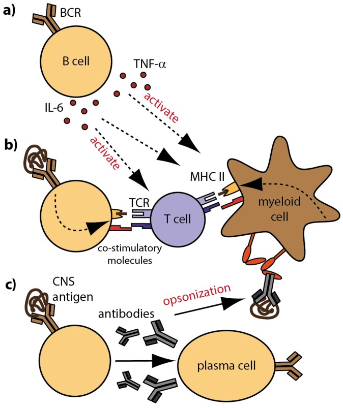
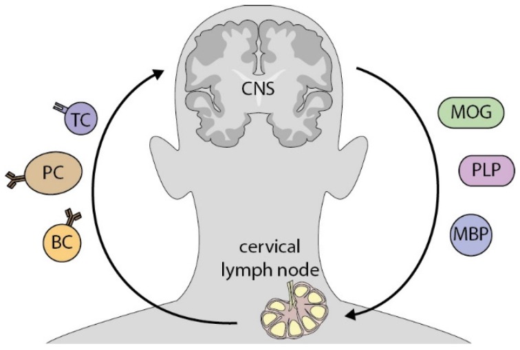
In central nervous system (CNS) demyelinating disorders, such as multiple sclerosis (MS), neuromyelitis optica (NMO) and related NMO-spectrum disorders (NMO-SD), a pathogenic role for antibodies is primarily projected into enhancing ongoing CNS inflammation by directly binding to target antigens within the CNS. This scenario is supported at least in part, by antibodies in conjunction with complement activation in the majority of MS lesions and by deposition of anti-aquaporin-4 (AQP-4) antibodies in areas of astrocyte loss in patients with classical NMO. A currently emerging subgroup of AQP-4 negative NMO-SD patients expresses antibodies against myelin oligodendrocyte glycoprotein (MOG), again suggestive of their direct binding to CNS myelin. However, both known entities of anti-CNS antibodies, anti-AQP-4- as well as anti-MOG antibodies, are predominantly found in the serum, which raises the questions why and how a humoral response against CNS antigens is raised in the periphery, and in a related manner, what pathogenic role these antibodies may exert outside the CNS. In this regard, recent experimental and clinical evidence suggests that peripheral CNS-specific antibodies may indirectly activate peripheral CNS-autoreactive T cells by opsonization of otherwise unrecognized traces of CNS antigen in peripheral compartments, presumably drained from the CNS by its newly recognized lymphatic system. In this review, we will summarize all currently available data on both possible roles of antibodies in CNS demyelinating disorders, first, directly enhancing damage within the CNS, and second, promoting a peripheral immune response against the CNS. By elaborating on the latter scenario, we will develop the hypothesis that peripheral CNS-recognizing antibodies may have a powerful role in initiating acute flares of CNS demyelinating disease and that these humoral responses may represent a therapeutic target in its own right.

Keywords: CNS-draining lymphatics; aquaporin-4; autoantibody; central nervous system; multiple sclerosis; myelin oligodendrocyte glycoprotein; neuromyelitis optica; opsonization.


Conflict of interest statement

The authors declare no conflict of interest.

Figures

Figure 1
Figure 1
Cellular and molecular B cell properties in MS; (a) B cells modulate the activation and differentiation of immune cells by secretion of pro- and anti-inflammatory cytokines; (b) Antigen-specific B cells recognize CNS antigen via their BCR and internalize, process and present linearized antigens to responding T cells. Ligation of co-stimulatory molecules and secretion of pro-inflammatory cytokines foster the generation of effector T cells; (c) B cells differentiate into antibody-producing plasma cells. Secreted CNS-reactive antibodies that reach the CNS contribute to demyelination and inflammation by complement-mediated cytotoxicity. In the periphery, opsonization of rare CNS antigen by antibodies fosters the generation of auto-reactive T cells; Fc receptors on myeloid APC recognize antibody-antigen complexes and trigger internalization, processing and presentation of opsonized antigen to responding T cells. Definitions: APC = antigen-presenting cells; BCR = B cell receptor; CNS = central nervous system.
Figure 2
Figure 2
CNS antigens potentially activate peripheral immune cells in CNS-draining lymph nodes. Cerebrospinal fluid, which occasionally contains CNS antigen, such as myelin components, is drained by lymphatic vessels into deep cervical lymph nodes. There, antigen is encountered and processed by professional APC and subsequently presented to autoreactive antigen-specific T cells. By this interaction, immune cells are activated and in turn, migrate to the CNS, where they contribute to inflammation. Definitions: MOG = myelin oligodendrocyte glycoprotein; PLP = proteolipid-Protein 1; MBP = myelin basic protein; TC = T cell, PC = plasma cell; BC = B cell.

References

    1. Kinzel S., Weber M.S.B. Cell-Directed Therapeutics in Multiple Sclerosis: Rationale and Clinical Evidence. CNS Drug. 2016;30:1137–1148. doi: 10.1007/s40263-016-0396-6. - DOI - PubMed
    1. Bennett J.L., O’Connor K.C., Bar-Or A., Zamvil S.S., Hemmer B., Tedder T.F., von Büdingen H.C., Stuve O., Yeaman M.R., Smith T.J., et al. B lymphocytes in neuromyelitis optica. Neurol. Neuroimmunol. Neuroinflam. 2015;2:e104. doi: 10.1212/NXI.0000000000000104. - DOI - PMC - PubMed
    1. Hauser S.L., Bar-Or A., Comi G., Giovannoni G., Hartung H.P., Hemmer B., Lublin F., Montalban X., Rammohan K.W., Selmaj K., et al. Ocrelizumab versus Interferon Beta-1a in Relapsing Multiple Sclerosis. N. Engl. J. Med. 2017;376:221–234. doi: 10.1056/NEJMoa1601277. - DOI - PubMed
    1. Montalban X., Hauser S.L., Kappos L., Arnold D.L., Bar-Or A., Comi G., de Seze J., Giovannoni G., Hartung H.-P., Hemmer B., et al. Ocrelizumab versus Placebo in Primary Progressive Multiple Sclerosis. N. Engl. J. Med. 2017;376:209–220. doi: 10.1056/NEJMoa1606468. - DOI - PubMed
    1. Cree B.A., Lamb S., Morgan K., Chen A., Waubant E., Genain C. An open label study of the effects of rituximab in neuromyelitis optica. Neurology. 2005;64:1270–1272. doi: 10.1212/01.WNL.0000159399.81861.D5. - DOI - PubMed

LinkOut - more resources