. 2018 Jan 2;12(1):37-46.
doi: 10.1080/19336918.2017.1313382. Epub 2017 Jun 29.

PHLDA1 (pleckstrin homology-like domain, family A, member 1) knockdown promotes migration and invasion of MCF10A breast epithelial cells

Affiliations

PHLDA1 (pleckstrin homology-like domain, family A, member 1) knockdown promotes migration and invasion of MCF10A breast epithelial cells

Naieli Bonatto et al. Cell Adh Migr. .

Abstract

PHLDA1 (pleckstrin homology-like domain, family A, member 1) is a multifunctional protein that plays distinct roles in several biological processes including cell death and therefore its altered expression has been identified in different types of cancer. Progressively loss of PHLDA1 was found in primary and metastatic melanoma while its overexpression was reported in intestinal and pancreatic tumors. Previous work from our group showed that negative expression of PHLDA1 protein was a strong predictor of poor prognosis for breast cancer disease. However, the function of PHLDA1 in mammary epithelial cells and the tumorigenic process of the breast is unclear. To dissect PHLDA1 role in human breast epithelial cells, we generated a clone of MCF10A cells with stable knockdown of PHLDA1 and performed functional studies. To achieve reduced PHLDA1 expression we used shRNA plasmid transfection and then changes in cell morphology and biological behavior were assessed. We found that PHLDA1 downregulation induced marked morphological alterations in MCF10A cells, such as changes in cell-to-cell adhesion pattern and cytoskeleton reorganization. Regarding cell behavior, MCF10A cells with reduced expression of PHLDA1 showed higher proliferative rate and migration ability in comparison with control cells. We also found that MCF10A cells with PHLDA1 knockdown acquired invasive properties, as evaluated by transwell Matrigel invasion assay and showed enhanced colony-forming ability and irregular growth in low attachment condition. Altogether, our results indicate that PHLDA1 downregulation in MCF10A cells leads to morphological changes and a more aggressive behavior.

Keywords: MCF10A; PHLDA1; aggressive phenotype; breast cancer; invasion; mammary epithelial cells; migration.


Figures

Figure 1.
Figure 1.
PHLDA1 protein expression and morphological features of MCF10A control and PHLDA1 knockdown cells. (A) Western blot analysis of PHLDA1 expression in MCF10A cells. First lane, MCF10A control cells (Ctrl); Lanes 2 and 3, MCF10A shPHLDA1 #1 and #2 sub-clones, respectively. (B) Phase-contrast images showing the morphological characteristics of MCF10A control and PHLDA1 knockdown cells grown in monolayer. Confluent and sub-confluent MCF10A control cells exhibiting typical morphology with lamellipodia often observed at the edges of clusters at sub-confluence. At confluence, MCF10A PHLDA1 knockdown cells assumed typical cobblestone morphology similar to control cells; at sub-confluence, cells exhibited lamellipodium more extensive than control cells (black arrows).
Figure 2.
Figure 2.
PHLDA1 knockdown increases proliferation and enhances mammosphere formation in MCF10A cells. (A) Increased proliferation rate in knockdown PHLDA1 MCF10A cells as compared with control cells. The cell proliferation rate was determined using a CyQUANT cell proliferation assay after 24, 48, 72 and 96 hours. Curve fit analysis was performed using GraphPad Prism software for nonlinear regression. (Left) Control cells versus shPHLDA1 #1 sub-clone, P < 0.01. (Right) Control cells vs. shPHLDA1 #2 sub-clone, P < 0.05. (B) MCF10A control and PHLDA1 knockdown mammospheres characteristics. (a) MCF10A control and (c) PHLDA1 knockdown cells were seeded at single-cell density in low attachment plates and mammosphere were allowed to develop for 9 days, the mean size of mammospheres for each lineage was plotted (b) and the total number of mammospheres were counted (d) and plotted with mean ± s.e.m (ns, unpaired t test).
Figure 3.
Figure 3.
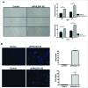
PHLDA1 knockdown enhances migration and promotes invasion of MCF10A cells. (A) Wound-healing assay shows that PHLDA1 knockdown significantly increases cell mobility. (Left) representative photomicrographs of cells. (Right) The bar graph shows the quantitative data for wound closure after 18 and 24 hours. Data represents mean ± s.e.m. **P < 0.01; ***p < 0.001 by 2-way ANOVA test with Bonferroni correction. (B) PHLDA1 knockdown promotes invasion of MCF10A cells. (Upper) Representative photomicrographs of cells that have invaded, stained with DAPI. (Lower) Graph represents mean ± s.e.m. for the number of cells that have invaded/field in transwells from 3 independent experiments **P < 0.01 (unpaired t test).
Figure 4.
Figure 4.
PHLDA1 knockdown enhances the ability of MCF10A cells to form colonies. (A) Representative images taken from 6 well plates showing colonies of MCF10A control cells, shPHLDA1 #1 and #2 cells. (B) Bar graph showing number of colonies after 8 d in culture. Data are expressed as mean ± s.e.m. *P < 0.05 (unpaired t test). (C) Morphological differences between MCF10A control and PHLDA1 knockdown cells colonies stained with crystal violet. (Left) Representative photomicrograph of control cells. (Right) Representative photomicrograph of PHLDA1 knockdown cells.
Figure 5.
Figure 5.
PHLDA1 downregulation induces morphological changes in MCF10A cells. Actin staining revealed morphological differences between Control and shPHLDA1 cells. (A) MCF10A PHLDA1 knockdown cells display highly actin-rich membrane projections. Representative photomicrographs of cells immunostained with actin primary antibody and Alexa-Fluor 488 secundary antibody (green) and nuclei counterstained with DAPI. (B) Contrast between the tight cell-cell contact of control cells with the loose cell-cell contact of shPHLDA1 cells. Representative photomicrographs of cells stained with actin antibody (green), rhodamin phalloidin (red) and nuclei counterstained with DAPI. Scale bar: 20 μm.

References

    1. Nagai MA. Pleckstrin homology-like domain, family A, member 1 (PHLDA1) and cancer. Biomed Rep 2016; 4(3):275-81; PMID:26998263 - PMC - PubMed
    1. Oberst MD, Beberman SJ, Zhao L, Yin JJ, Ward Y, Kelly K. TDAG51 is an ERK signaling target that opposes ERK-mediated HME16C mammary epithelial cell transformation. BMC Cancer 2008; 8:189; PMID:18597688; https://doi.org/10.1186/1471-2407-8-189 - DOI - PMC - PubMed
    1. Johnson EO, Chang KH, de Pablo Y, Ghosh S, Mehta R, Badve S, Shah K. PHLDA1 is a crucial negative regulator and effector of Aurora A kinase in breast cancer. J Cell Sci 2011; 124(Pt 16):2711-22; PMID:21807936; https://doi.org/10.1242/jcs.084970 - DOI - PubMed
    1. Moad AI, Muhammad TS, Oon CE, Tan ML. Rapamycin induces apoptosis when autophagy is inhibited in T-47D mammary cells and both processes are regulated by Phlda1. Cell Biochem Biophys 2013; 66(3):567-87; PMID:23300026; https://doi.org/10.1007/s12013-012-9504-5 - DOI - PubMed
    1. Li G, Wang X, Hibshoosh H, Jin C, Halmos B. Modulation of ErbB2 blockade in ErbB2-positive cancers: the role of ErbB2 Mutations and PHLDA1. PLoS One 2014; 9(9):e106349; PMID:25238247; https://doi.org/10.1371/journal.pone.0106349 - DOI - PMC - PubMed

Publication types

MeSH terms

LinkOut - more resources