

Primary CNS Lymphoma
- PMID: 28640701
- PMCID: PMC5516483
- DOI: 10.1200/JCO.2017.72.7602
Primary CNS Lymphoma
Abstract
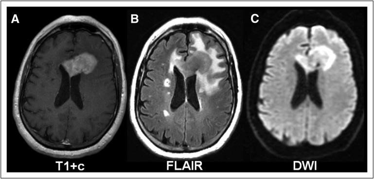
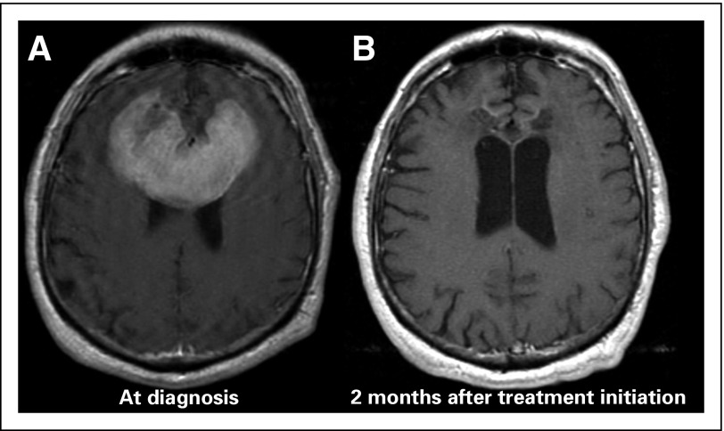
Primary CNS lymphoma (PCNSL) is a rare form of extranodal non-Hodgkin lymphoma that is typically confined to the brain, eyes, and cerebrospinal fluid without evidence of systemic spread. The prognosis of patients with PCNSL has improved during the last decades with the introduction of high-dose methotrexate. However, despite recent progress, results after treatment are durable in half of patients, and therapy can be associated with late neurotoxicity. PCNSL is an uncommon tumor, and only four randomized trials and one phase III trial have been completed so far, all in the first-line setting. To our knowledge, no randomized trial has been conducted for recurrent/refractory disease, leaving many questions unanswered about optimal first-line and salvage treatments. This review will give an overview of the presentation, evaluation, and treatment of immunocompetent patients with PCNSL.
Figures
References
-
- O’Neill BP, Decker PA, Tieu C, et al. : The changing incidence of primary central nervous system lymphoma is driven primarily by the changing incidence in young and middle-aged men and differs from time trends in systemic diffuse large B-cell non-Hodgkin’s lymphoma. Am J Hematol 88:997-1000, 2013 - PMC - PubMed
-
- Bataille B, Delwail V, Menet E, et al. : Primary intracerebral malignant lymphoma: Report of 248 cases. J Neurosurg 92:261-266, 2000 - PubMed
-
- Grimm SA, Pulido JS, Jahnke K, et al. : Primary intraocular lymphoma: An International Primary Central Nervous System Lymphoma Collaborative Group Report. Ann Oncol 18:1851-1855, 2007 - PubMed
-
- Batchelor T, Carson K, O’Neill A, et al. : Treatment of primary CNS lymphoma with methotrexate and deferred radiotherapy: A report of NABTT 96-07. J Clin Oncol 21:1044-1049, 2003 - PubMed
Publication types
MeSH terms
Grants and funding
LinkOut - more resources
Full Text Sources
Other Literature Sources
