Review
. 2017 Jul 20;35(21):2439-2449.
doi: 10.1200/JCO.2017.72.7511. Epub 2017 Jun 22.

Response Assessment in Neuro-Oncology Clinical Trials

Affiliations
Review

Response Assessment in Neuro-Oncology Clinical Trials

Patrick Y Wen et al. J Clin Oncol. .

Abstract

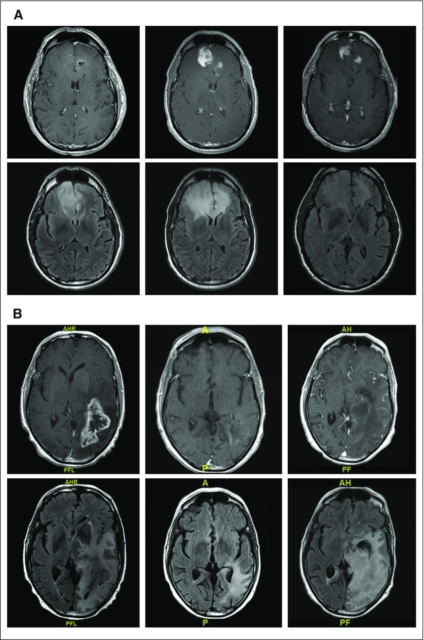
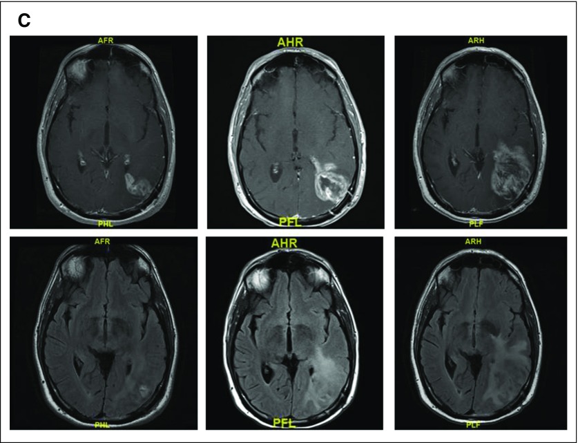
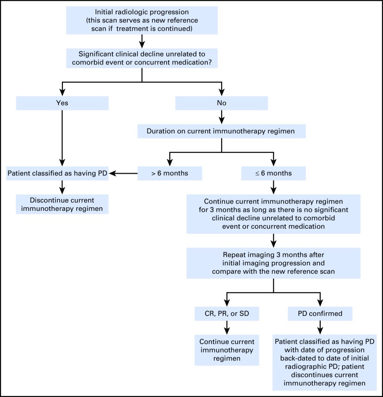
Development of novel therapies for CNS tumors requires reliable assessment of response and progression. This requirement has been particularly challenging in neuro-oncology for which contrast enhancement serves as an imperfect surrogate for tumor volume and is influenced by agents that affect vascular permeability, such as antiangiogenic therapies. In addition, most tumors have a nonenhancing component that can be difficult to accurately quantify. To improve the response assessment in neuro-oncology and to standardize the criteria that are used for different CNS tumors, the Response Assessment in Neuro-Oncology (RANO) working group was established. This multidisciplinary international working group consists of neuro-oncologists, medical oncologists, neuroradiologists, neurosurgeons, radiation oncologists, neuropsychologists, and experts in clinical outcomes assessments, working in collaboration with government and industry to enhance the interpretation of clinical trials. The RANO working group was originally created to update response criteria for high- and low-grade gliomas and to address such issues as pseudoresponse and nonenhancing tumor progression from antiangiogenic therapies, and pseudoprogression from radiochemotherapy. RANO has expanded to include working groups that are focused on other tumors, including brain metastases, leptomeningeal metastases, spine tumors, pediatric brain tumors, and meningiomas, as well as other clinical trial end points, such as clinical outcomes assessments, seizures, corticosteroid use, and positron emission tomography imaging. In an effort to standardize the measurement of neurologic function for clinical assessment, the Neurologic Assessment in Neuro-Oncology scale was drafted. Born out of a workshop conducted by the Jumpstarting Brain Tumor Drug Development Coalition and the US Food and Drug Administration, a standardized brain tumor imaging protocol now exists to reduce variability and improve reliability. Efforts by RANO have been widely accepted and are increasingly being used in neuro-oncology trials, although additional refinements will be needed.


Figures

Fig 1.
Fig 1.
(A) T1 postcontrast and fluid-attenuated inversion recovery (FLAIR) images before (left), 2 months after (middle), and 3 months (right) after completing radiation and concurrent temozoloimde in a patient with a newly diagnosed glioblastoma (GBM). The extent of enhancement initially increases, but then improves with time to suggest that the initial post-treatment changes represented pseudoprogression. (B) T1 postcontrast and FLAIR images before (left), after one cycle (middle), and after five cycles (right) of bevacizumab in a patient with recurrent GBM. Although enhancement remains nonmeasurable after five cycles of treatment, the extent of FLAIR only continues to expand in this case of nonenhancing disease progression. (C) T1 postcontrast and FLAIR images before (left), 1 month after (middle), and 3 months (right) after completing radiation and concurrent nivolumab in a patient with a newly diagnosed GBM. Tumor resection pathology revealed almost all treatment effect.
Fig 1.
Fig 1.
(A) T1 postcontrast and fluid-attenuated inversion recovery (FLAIR) images before (left), 2 months after (middle), and 3 months (right) after completing radiation and concurrent temozoloimde in a patient with a newly diagnosed glioblastoma (GBM). The extent of enhancement initially increases, but then improves with time to suggest that the initial post-treatment changes represented pseudoprogression. (B) T1 postcontrast and FLAIR images before (left), after one cycle (middle), and after five cycles (right) of bevacizumab in a patient with recurrent GBM. Although enhancement remains nonmeasurable after five cycles of treatment, the extent of FLAIR only continues to expand in this case of nonenhancing disease progression. (C) T1 postcontrast and FLAIR images before (left), 1 month after (middle), and 3 months (right) after completing radiation and concurrent nivolumab in a patient with a newly diagnosed GBM. Tumor resection pathology revealed almost all treatment effect.
Fig A1.
Fig A1.
Response Assessment in Neuro-Oncology treatment algorithm for the assessment of progressive imaging findings in neuro-oncology patients treated with immunotherapy. CR, complete response; PD, progressive disease; PR, partial response; SD, stable disease. Reprinted from Okada et al with permission from Elsevier.

References

    1. Miller JJ, Wen PY: Emerging targeted therapies for glioma. Expert Opin Emerg Drugs 21:441-452, 2016 - PubMed
    1. Wen PY, Cloughesy TF, Ellingson BM, et al. : Report of the Jumpstarting Brain Tumor Drug Development Coalition and FDA clinical trials neuroimaging endpoint workshop (January 30, 2014, Bethesda MD). Neuro-oncol 16:vii36-vii47, 2014. (suppl 7) - PMC - PubMed
    1. van den Bent MJ, Vogelbaum MA, Wen PY, et al. : End point assessment in gliomas: Novel treatments limit usefulness of classical Macdonald’s Criteria. J Clin Oncol 27:2905-2908, 2009 - PMC - PubMed
    1. Wen PY, Macdonald DR, Reardon DA, et al. : Updated response assessment criteria for high-grade gliomas: Response assessment in neuro-oncology working group. J Clin Oncol 28:1963-1972, 2010 - PubMed
    1. Chang S, Wen P, Vogelbaum M, et al. : Response Assessment in Neuro-Oncology (RANO): More than imaging criteria for malignant glioma. Neuro-Oncol Pract 2:205-209, 2015 - PMC - PubMed

MeSH terms