. 2017 Oct;143(2):236-243.
doi: 10.1111/jnc.14110. Epub 2017 Aug 4.

The interaction between progranulin and prosaposin is mediated by granulins and the linker region between saposin B and C

Affiliations

The interaction between progranulin and prosaposin is mediated by granulins and the linker region between saposin B and C

Xiaolai Zhou et al. J Neurochem. 2017 Oct.

Abstract

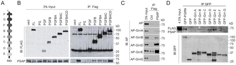
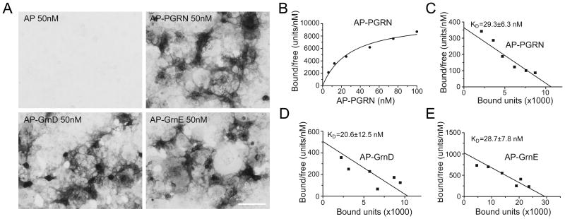
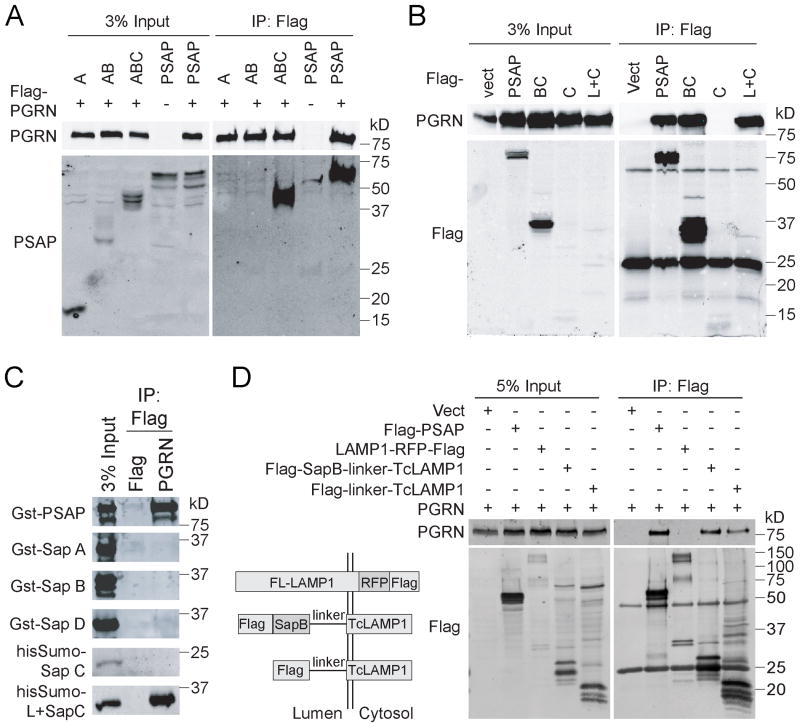
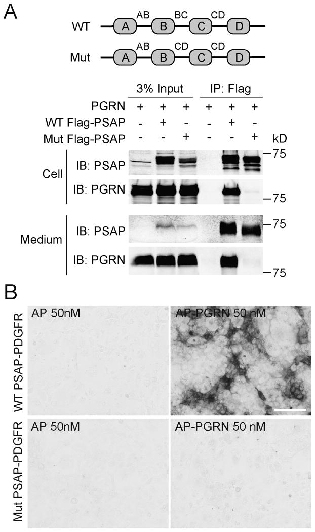
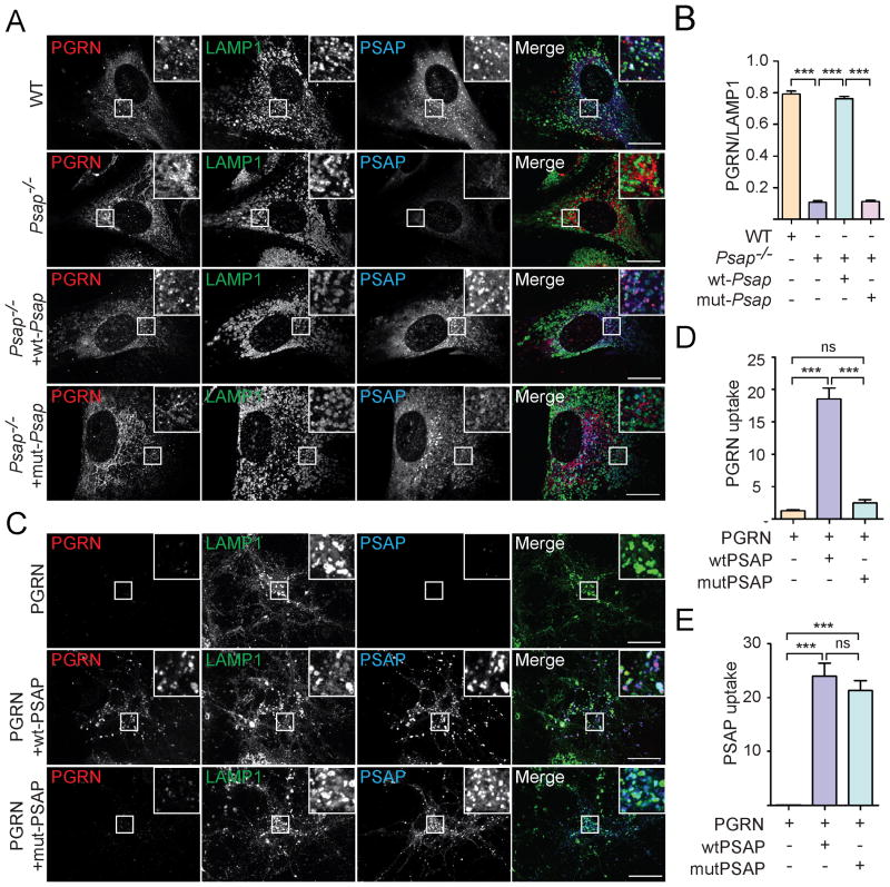
The frontotemporal lobar degeneration (FTLD) protein progranulin (PGRN) is essential for proper lysosomal function. PGRN localizes in the lysosomal compartment within the cell. Prosaposin (PSAP), the precursor of lysosomal saposin activators (saposin A, B, C, D), physically interacts with PGRN. Previously, we have shown that PGRN and PSAP facilitate each other's lysosomal trafficking. Here, we report that the interaction between PSAP and PGRN requires the linker region of saposin B and C (BC linker). PSAP protein with the BC linker mutated, fails to interact with PGRN and deliver PGRN to lysosomes in the biosynthetic and endocytic pathways. On the other hand, PGRN interacts with PSAP through multiple granulin motifs. Granulin D and E bind to PSAP with similar affinity as full-length PGRN. Read the Editorial Comment for this article on page 154.

Keywords: frontotemporal lobar degeneration; lysosomal storage diseases; lysosome; neuronal ceroid lipofuscinosis; progranulin; prosaposin.


Conflict of interest statement

Conflict of interest: none.

Figures

Figure 1
Figure 1. PSAP interacts with granulin motifs
(A) Domain structure of human PGRN (aa 1-593). (B) HEK293T cells were co-transfected with FLAG tagged PGRN truncation constructs and untagged human PSAP as indicated. Cells were lysed two days after transfection and the lysates were immunoprecipitated with anti-FLAG antibodies. The IP products were analyzed by Western blot using anti-FLAG and anti-PSAP antibodies (RRID:AB_2172462). (C) Conditioned medium from HEK293T expressing different AP-fusion proteins were incubated with FLAG beads only or FLAG beads with FLAG-PSAP recombinant proteins. The amount of AP proteins co-immunoprecipitated with FLAG-PSAP was analyzed by Western blot. (D) Conditioned medium from HEK293T expressing different GFP-fusion proteins were mixed with 1μg purified recombinant FLAG-PSAP. Immunoprecipitations were then performed with anti-GFP antibodies and the IP products were analyzed by Western blot.
Figure 2
Figure 2. Granulin D and E bind to PSAP with similar affinity as full length PGRN
(A) Conditioned medium containing AP, AP-PGRN, AP-Grn D, or AP-Grn E were incubated with COS-7 cells transfected with PSAP fused to PDGFR. Cells were fixed and AP binding was visualized with AP substrates. Scale bar=100μm. (B) AP–PGRN binding to PSAP-PDGFR expressing COS-7 cells measured as a function of AP–PGRN concentration. (C-E) Scatchard plot of AP-PGRN (C), AP-GRN D (D) or AP-GRN E (E) binding to PSAP-PDGFR expressing COS-7 cells. KD, mean ± sem, n = 4.
Figure 3
Figure 3. PSAP interacts with PGRN through the BC linker
(A) HEK293T cells were co-transfected with FLAG tagged PGRN and full length his-PSAP or PSAP truncation mutants as indicated. Cells were lysed two days after transfection and the lysates were immunoprecipitated with anti-FLAG antibodies. The IP products were analyzed by Western blot using anti-FLAG and anti-PSAP antibodies (RRID:AB_2172462). (B) HEK293T cells were co-transfected with untagged PGRN and FLAG tagged PSAP fragments as indicated. Cells were lysed two days after transfection and the lysates were immunoprecipitated with anti-FLAG antibodies. (C) Recombinant Gst- or his-sumo tagged saposins purified from bacteria were incubated with FLAG beads only or FLAG beads with FLAG-PGRN recombinant proteins. The amount of saposin co-immunoprecipitated with FLAG-PGRN was analyzed by Western blot. (D) HEK293T cells were co-transfected with untagged PGRN and FLAG tagged full-length PSAP, full-length LAMP1, saposin B with BC linker fused with transmembrane and cytoplasmic domain of LAMP1 (Tc-LAMP1) and BC linker fused with Tc-LAMP1 as indicated. Cells were lysed two days after transfection and the lysates were immunoprecipitated with anti-FLAG antibodies and immunoblotted with anti-PGRN and anti-FLAG antibodies.
Figure 4
Figure 4. PSAP mutant with the BC linker replaced failed to interact with PGRN
(A) HEK293T cells were co-transfected with FLAG tagged wild type (WT) or mutant (mut) PSAP and untagged human PGRN as indicated. Media was collected and cells were lysed three days after transfection. The lysates and media were separately immunoprecipitated with anti-FLAG antibodies. (B) Conditioned medium containing AP or AP-PGRN were incubated with COS-7 cells transfected with WT or mutant PSAP fused to PDGFR. Cells were fixed and AP binding were visualized with AP substrates. Scale bar=100μm.
Figure 5
Figure 5. PSAP mutants fail to deliver PGRN to lysosomes
(A) PSAP-/- mouse fibroblasts were infected with lentivirus expressing WT or mutant PSAP with the BC linker replaced by CD linker. Cells were fixed and stained with rabbit anti-human saposin B, sheep anti-mouse PGRN and rat anti-LAMP1 antibodies. Scale bar=10 μm. (B) The colocalization of PGRN with lysosomal marker LAMP1 in (A) were quantified using Image J. n=3, ***, p<0.001. (C) Grn-/- cortical neurons were incubated with C-terminally FLAG his tagged human PGRN which does not bind to sortilin (5ug/ml) together with recombinant WT PSAP or mutant PSAP with the BC linker replaced by CD linker (5ug/ml). 12 hours later, cells were fixed and stained with rabbit anti-human saposin B, goat anti-human PGRN and rat anti-LAMP1 antibodies. Scale bar=10 μm. (D, E) The endocytosed PGRN (D) and PSAP (E) for the experiment in (C) were quantified by using Image J. n=3; ns, no significance; ***, p<0.001. One way ANOVA.

References

    1. Ahmed Z, Mackenzie IR, Hutton ML, Dickson DW. Progranulin in frontotemporal lobar degeneration and neuroinflammation. J Neuroinflammation. 2007;4:7. - PMC - PubMed
    1. Almeida MR, Macario MC, Ramos L, Baldeiras I, Ribeiro MH, Santana I. Portuguese family with the co-occurrence of frontotemporal lobar degeneration and neuronal ceroid lipofuscinosis phenotypes due to progranulin gene mutation. Neurobiol Aging 2016 - PubMed
    1. Arai T, Hasegawa M, Akiyama H, Ikeda K, Nonaka T, Mori H, Mann D, Tsuchiya K, Yoshida M, Hashizume Y, et al. TDP-43 is a component of ubiquitin-positive tau-negative inclusions in frontotemporal lobar degeneration and amyotrophic lateral sclerosis. Biochem Biophys Res Commun. 2006;351:602–611. - PubMed
    1. Baker M, Mackenzie IR, Pickering-Brown SM, Gass J, Rademakers R, Lindholm C, Snowden J, Adamson J, Sadovnick AD, Rollinson S, et al. Mutations in progranulin cause tau-negative frontotemporal dementia linked to chromosome 17. Nature. 2006;442:916–919. - PubMed
    1. Bateman A, Bennett HP. The granulin gene family: from cancer to dementia. Bioessays. 2009;31:1245–1254. - PubMed