. 2017 Jun 21;94(6):1142-1154.e6.
doi: 10.1016/j.neuron.2017.06.008.

Enhanced Functional Genomic Screening Identifies Novel Mediators of Dual Leucine Zipper Kinase-Dependent Injury Signaling in Neurons

Affiliations

Enhanced Functional Genomic Screening Identifies Novel Mediators of Dual Leucine Zipper Kinase-Dependent Injury Signaling in Neurons

Derek S Welsbie et al. Neuron. .

Abstract

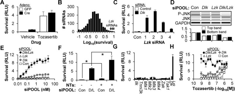
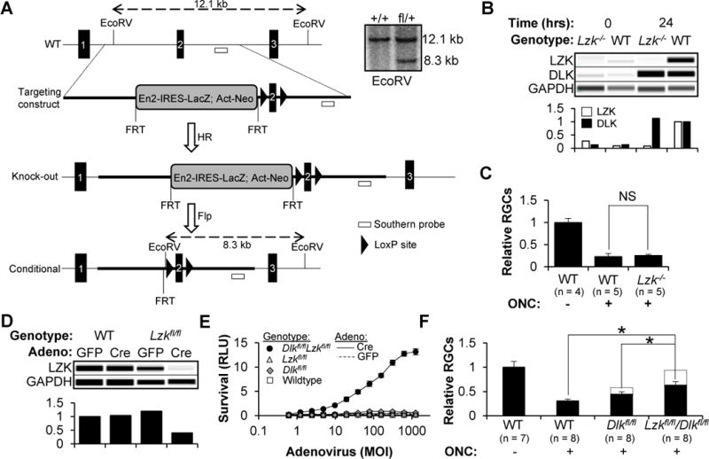
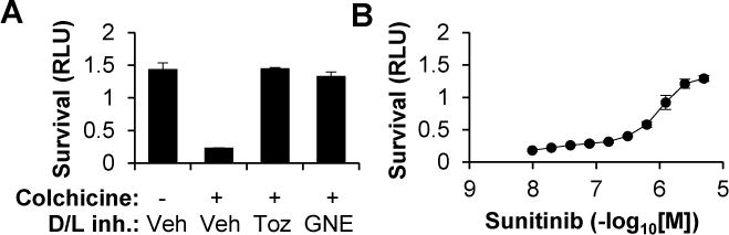
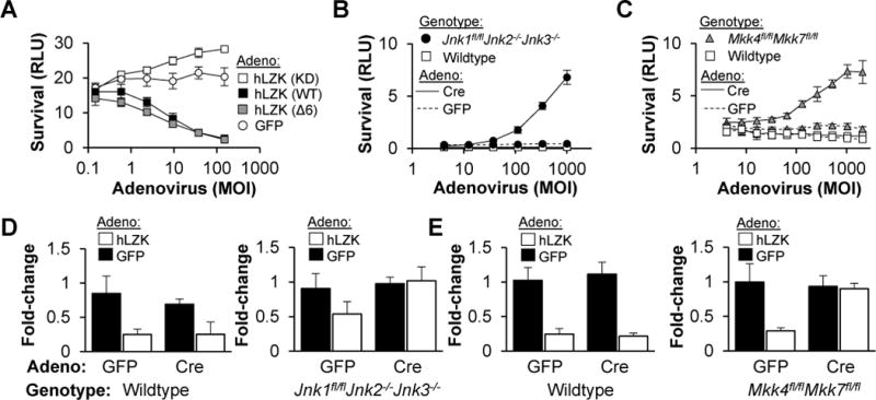
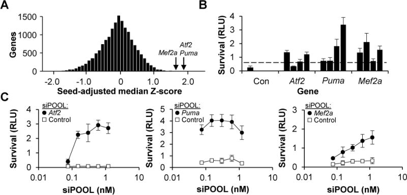
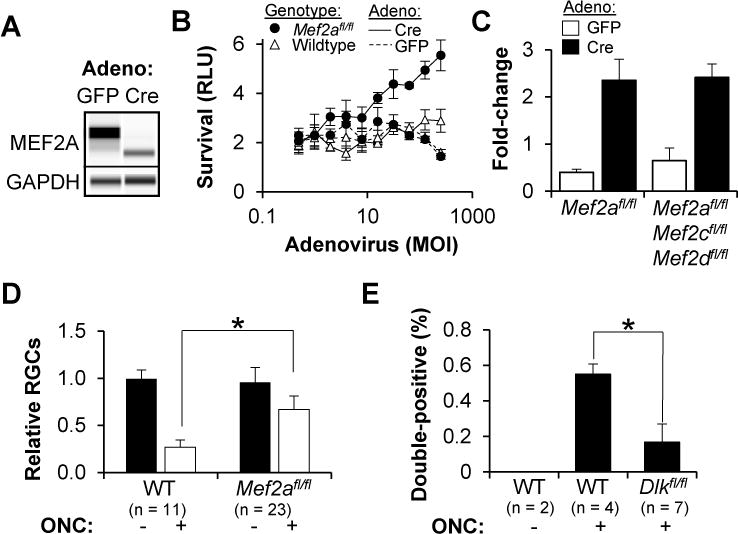
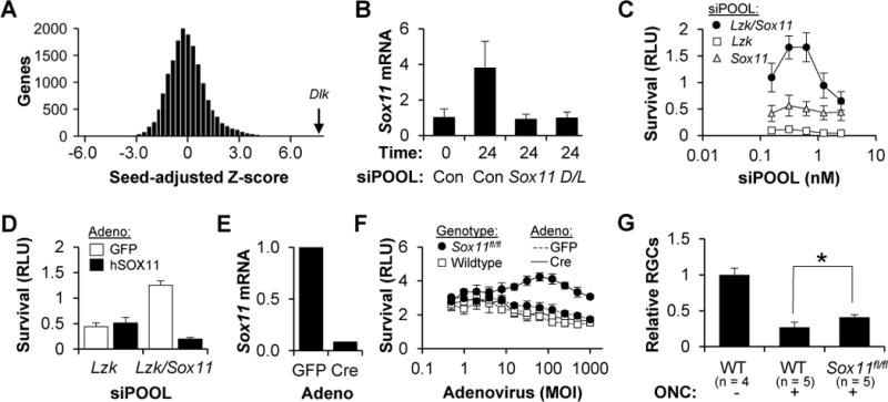
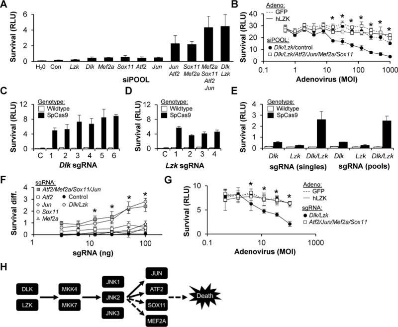
Dual leucine zipper kinase (DLK) has been implicated in cell death signaling secondary to axonal damage in retinal ganglion cells (RGCs) and other neurons. To better understand the pathway through which DLK acts, we developed enhanced functional genomic screens in primary RGCs, including use of arrayed, whole-genome, small interfering RNA libraries. Explaining why DLK inhibition is only partially protective, we identify leucine zipper kinase (LZK) as cooperating with DLK to activate downstream signaling and cell death in RGCs, including in a mouse model of optic nerve injury, and show that the same pathway is active in human stem cell-derived RGCs. Moreover, we identify four transcription factors, JUN, activating transcription factor 2 (ATF2), myocyte-specific enhancer factor 2A (MEF2A), and SRY-Box 11 (SOX11), as being the major downstream mediators through which DLK/LZK activation leads to RGC cell death. Increased understanding of the DLK pathway has implications for understanding and treating neurodegenerative diseases.

Keywords: DLK (dual leucine zipper kinase); LZK (leucine zipper kinase); Neuroprotection; RGC (retinal ganglion cell); RNAi screen; cell death signaling; glaucoma.


Figures

Figure 1
Figure 1. Sensitized siRNA screening of the kinome identifies LZK as a mediator of RGC cell death in vitro
(A) Survival (±SD) of Dlkfl/fl RGCs, transduced with adenovirus and cultured with or without tozasertib (1 μM), two days after colchicine (1 μM) addition. (B) Histogram showing the normalized survival for all 1,869 siRNAs in the kinome library (transfected in the presence of Dlk siRNA). (C) Survival (±SD) of WT RGCs transfected with control or Dlk siRNA, and one of four independent Lzk siRNAs or the non-targeting control (Con), two days after colchicine (1 μM) addition. (D) Capillary-based immunoassay (top) and quantification (bottom) of JNK phosphorylation in WT RGCs, one day after transfection with siPOOLs. (E) Survival (±SD) of WT RGCs transfected with siPOOLs, two days after colchicine (1 μM) addition. (F) Survival (±SD) of WT RGCs transfected with siPOOLs, in the presence or absence of neurotrophins (NTs, 50 ng/mL BDNF, 5 ng/mL GDNF, 5 ng/mL CNTF), two days after colchicine (1 μM) addition. *P<0.05, Mann-Whitney U test. D/L, Dlk/Lzk (G) Survival (±SD) of WT RGCs, transfected with Dlk siRNA and either control siRNAs or one of four independent siRNAs targeting the other members of the MLK family of kinases, two days after colchicine (1 μM) addition. (H) Survival (±SD) of WT RGCs, transfected with siPOOLs and cultured in tozasertib, two days after colchicine (1 μM) addition.
Figure 2
Figure 2. RGCs with targeted deletion of Dlk and Lzk are resistant to axon injury-induced cell death in vitro and in vivo
(A) Approach used to generate constitutive and conditional Lzk knockout mice. Inset shows a Southern blot confirming the presence of a single targeting construct in the heterozygous animals. (B) Capillary-based immunoassay (top) and quantification (bottom) of DLK and LZK in RGCs, 0 or 24 hours after immunopanning injury. (C) FC-based quantification of surviving RGCs, normalized to the uninjured control (±SD), two weeks after ONC or sham surgery. NS, non-significant, Mann-Whitney U test. (D) Capillary-based immunoassay (top) and quantification (bottom) of LZK in RGCs one day after transduction with adenovirus. (E) Survival (±SD) of RGCs, transduced with adenovirus, two days after a colchicine challenge. Dlkfl/fl RGCs transduced with Cre have a 2–5-fold increase in survival over the controls. (F) FC-based quantification of surviving RGCs, normalized to the uninjured control (±SD), two weeks after ONC or sham surgery. All eyes were injected with 109 vg AAV2-Cre two weeks prior to the surgery. *P<0.05, Mann-Whitney U test.
Figure 3
Figure 3. Pharmacologic inhibition of DLK and LZK, including by sunitinib, an FDA-approved small-molecule inhibitor, promotes the survival of human ESC-derived RGCs
(G) Survival (±SD) of hESC-derived RGCs two days after a challenge with vehicle or colchicine (1 μM) in the presence of the DLK/LZK inhibitors tozasertib (1 μM), Genentech inhibitor 123 (0.1 μM) or a vehicle control. (H) Survival (±SD) of hESC-derived RGCs two days after a challenge with colchicine (1 μM) in the presence of increasing doses of sunitinib.
Figure 4
Figure 4. LZK kinase signaling triggers RGC cell death via the MKK4/7 and JNK1-3 kinase cascade
(A) Survival (±SD) of WT RGCs transfected with Dlk/Lzk siPOOL, two days after reconstitution of LZK signaling with adenovirus expressing mouse siRNA-resistant, human LZK cDNA or a GFP control (“LZK reconstitution assay”). (B–C) Survival (±SD) of RGCs, transduced with adenovirus, two days after colchicine (1 μM) addition. (D–E) Fold-change in survival (±SD) of RGCs, pretreated with adenovirus to eliminate JNK (D) or MKK4/7 (E) signaling, in response to adenovirus (MOI 400) to reconstitute LZK signaling.
Figure 5
Figure 5. Whole-genome siRNA screen identifies ATF2, PUMA and MEF2A as mediators of RGC cell death
(A) Histogram showing the normalized, seed-adjusted survival for the median survival-promoting siRNA targeting each of the 17,575 genes in the whole-genome library. (B) Survival (±SD) of WT RGCs, transfected with one of four independent siRNAs targeting Atf2, Puma or Mef2a or the non-targeting control, two days after colchicine addition. Dashed line shows the threshold of survival greater than 3SD from the negative control. (C) Survival (±SD) of WT RGCs, transfected with siPOOLs, two days after colchicine (1 μM) addition.
Figure 6
Figure 6. RGCs with a targeted disruption of the transcriptional regulatory domain of MEF2A are partially resistant to axon injury-induced cell death in vivo
(A) Capillary-based immunoassay of MEF2A in RGCs, two days after transduction with adenovirus. (B) Survival (±SD) of RGCs, transduced with adenovirus, two days after colchicine (1 μM) addition. (C) Fold-change in survival (±SD) of RGCs from adenoviral transduction (MOI 400), two days after colchicine (1 μM) addition. (D) FC-based quantification of surviving RGCs, normalized to the uninjured control (±SD), two weeks after ONC or sham surgery. All eyes were injected with 109 vg AAV2-Cre two weeks prior to the surgery. *P<0.05, Mann-Whitney U test. (E) FC-based quantification of TUBB3/phospho-S408 MEF2A double-positive cells, expressed as a percentage of total retina (±SD), two days after ONC or sham surgery. *P<0.05, Mann-Whitney U test.
Figure 7
Figure 7. Sensitized, whole-genome siRNA screen identifies SOX11 as a downstream mediator of RGC cell death
(A) Histogram showing the normalized, seed-adjusted survival for the siRNA minipool targeting each of the 16,698 genes in the whole-genome library (transfected in the presence of Lzk siPOOL). (B) QPCR assay for Sox11 mRNA, normalized to GAPDH levels (±SD), in WT RGCs transfected with siPOOLs, at the indicated time following immunopanning injury. (C) Survival (±SD) of WT RGCs, transfected with siPOOL, two days after colchicine (1 μM) addition. (D) Survival (±SD) of WT RGCs transfected with siPOOLs, two days after reconstitution of SOX11 signaling with an adenovirus expressing mouse siRNA-resistant human SOX11 cDNA or a GFP control. (E) QPCR assay of Sox11 mRNA, normalized to GAPDH levels, in Sox11fl/fl RGCs transduced with adenovirus. (F) Survival (±SD) of RGCs, transduced with adenovirus, two days after a colchicine (1 μM) addition. (G) FC-based quantification of surviving RGCs, normalized to the uninjured control (±SD), two weeks after ONC or sham surgery. All eyes were injected with 109 vg AAV2-Cre two weeks prior to the surgery. *P<0.05, Mann-Whitney U test.
Figure 8
Figure 8. DLK/LZK-dependent cell death is mediated by a set of four transcription factors: JUN, ATF2, SOX11 and MEF2A
(A) Survival (±SD) of WT RGCs transfected with siPOOL, two days after colchicine (1 μM) addition. (B) Survival (±SD) of WT RGCs transfected with siPOOL, two days after adenoviral transduction to reconstitute LZK signaling. *P<0.05 Mann-Whitney U test comparing absence/presence of transcription factor siPOOLs. (C–E) Survival (±SD) of RGCs, transfected with Lzk siPOOL and either tracrRNA or sgRNAs targeting Dlk (C), Dlk siPOOL and either tracrRNA or sgRNAs targeting Lzk (D) or pools versus individual sgRNAs targeting Dlk and Lzk (E). (F) Difference in survival (SpCas9-WT; ±SD) conferred by transfecting sgRNA targeting each of the four transcription factors, alone or in combination, and compared to negative control tracrRNA or positive control sgRNAs targeting Dlk/Lzk. *P<0.05 Mann-Whitney U test compared to control; no significant difference between Dlk/Lzk and transcription factor sgRNAs. (G) Survival (±SD) of SpCas9 RGCs, transfected with sgRNA, two days after adenoviral transduction to activate LZK signaling. *P<0.05 Mann-Whitney U test comparing Dlk/Lzk and transcription factor sgRNAs. (H) Proposed pathway for RGC cell death following axon injury. Dashed lines indicate that SOX11 and MEF2A are downstream of DLK/LZK, but not necessarily JNK1-3, and could be indirectly activated.

References

    1. Akhtar MW, Kim MS, Adachi M, Morris MJ, Qi X, Richardson JA, Bassel-Duby R, Olson EN, Kavalali ET, Monteggia LM. In vivo analysis of MEF2 transcription factors in synapse regulation and neuronal survival. PLoS One. 2012;7:e34863. - PMC - PubMed
    1. Ambacher KK, Pitzul KB, Karajgikar M, Hamilton A, Ferguson SS, Cregan SP. The JNK-and AKT/GSK3β-signaling pathways converge to regulate Puma induction and neuronal apoptosis induced by trophic factor deprivation. PLoS One. 2012;7:e46885. - PMC - PubMed
    1. Bounoutas A, Kratz J, Emtage L, Ma C, Nguyen KC, Chalfie M. Microtubule depolymerization in Caenorhabditis elegans touch receptor neurons reduces gene expression through a p38 MAPK pathway. Proc Natl Acad Sci U S A. 2011;108:3982–3987. - PMC - PubMed
    1. Buckingham BP, Inman DM, Lambert W, Oglesby E, Calkins DJ, Steele MR, Vetter ML, Marsh-Armstrong N, Horner PJ. Progressive ganglion cell degeneration precedes neuronal loss in a mouse model of glaucoma. J Neurosci. 2008;28:2735–2744. - PMC - PubMed
    1. Buehler E, Khan AA, Marine S, Rajaram M, Bahl A, Burchard J, Ferrer M. siRNA off-target effects in genome-wide screens identify signaling pathway members. Sci Rep. 2012;2:428. - PMC - PubMed

MeSH terms