. 2017 Aug 15;6(8):1229-1234.
doi: 10.1242/bio.026294.

Investigation of protein synthesis in Drosophila larvae using puromycin labelling

Affiliations

Investigation of protein synthesis in Drosophila larvae using puromycin labelling

Lisa P Deliu et al. Biol Open. .

Abstract

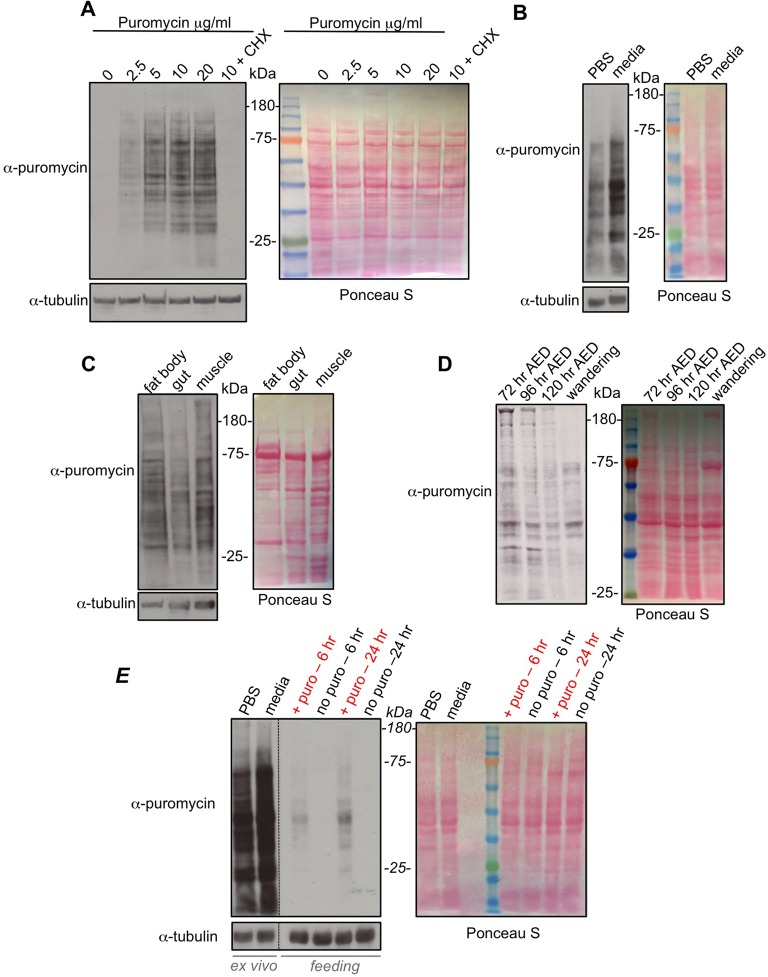
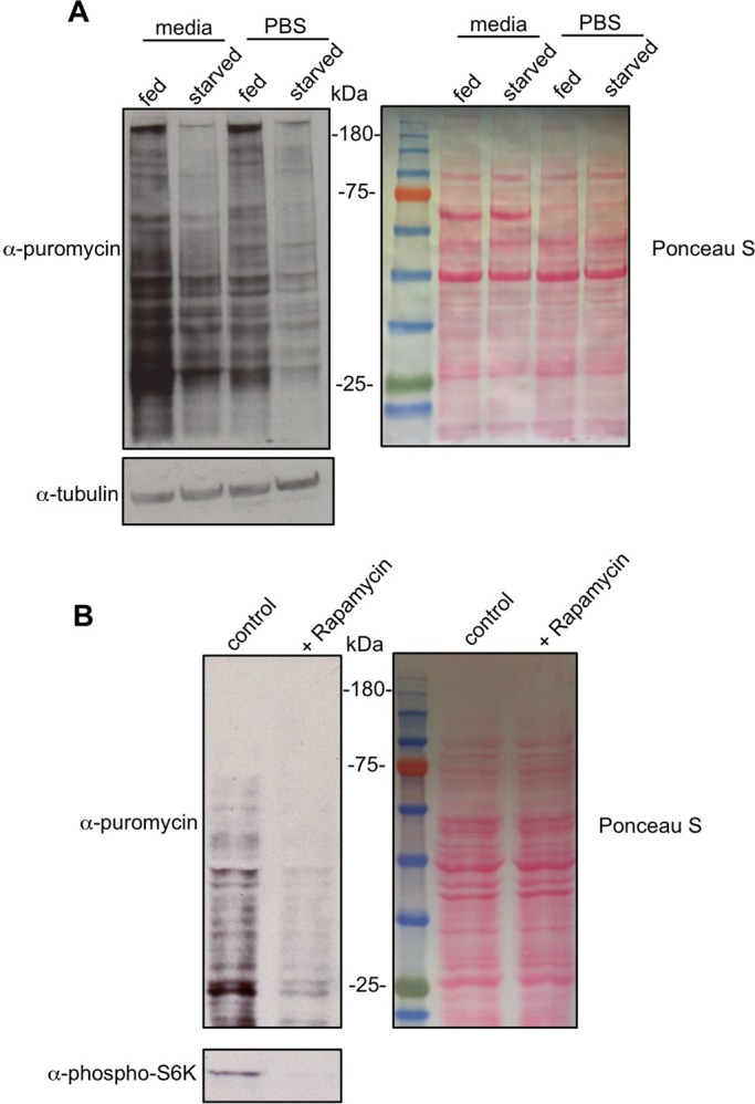
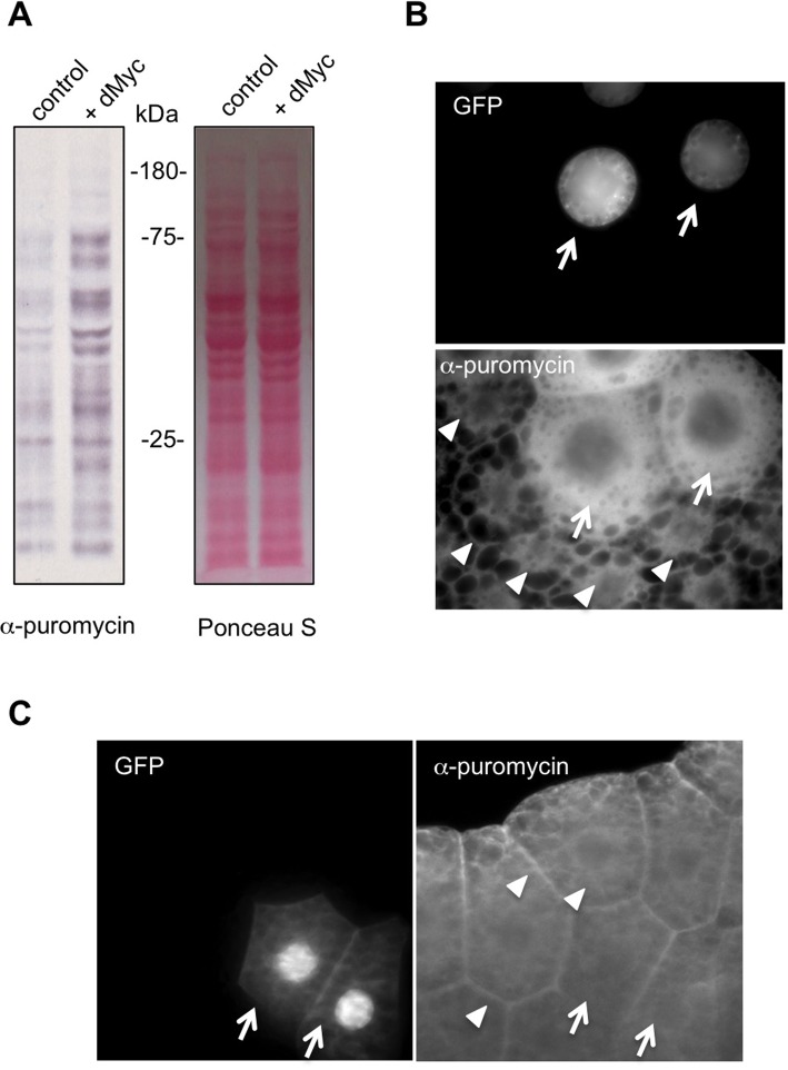
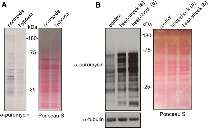
Translational control of gene expression is an important regulator of growth, homeostasis and aging in Drosophila The ability to measure changes in protein synthesis in response to genetic and environmental cues is therefore important in studying these processes. Here we describe a simple and cost-effective approach to assay protein synthesis in Drosophila larval cells and tissues. The method is based on the incorporation of puromycin into nascent peptide chains. Using an ex vivo approach, we label newly synthesized peptides in larvae with puromycin and then measure levels of new protein synthesis using an anti-puromycin antibody. We show that this method can detect changes in protein synthesis in specific cells and tissues in the larvae, either by immunostaining or western blotting. We find that the assay reliably detects changes in protein synthesis induced by two known stimulators of mRNA translation - the nutrient/TORC1 kinase pathway and the transcription factor dMyc. We also use the assay to describe how protein synthesis changes through larval development and in response to two environmental stressors - hypoxia and heat shock. We propose that this puromycin-labelling assay is a simple but robust method to detect protein synthesis changes at the levels of cells, tissues or whole body in Drosophila.

Keywords: Drosophila; Heat shock; Hypoxia; Nutrients; Protein synthesis; TOR kinase; dMyc; mRNA translation.


Conflict of interest statement

Competing interestsThe authors declare no competing or financial interests.

Figures

Fig. 1.
Fig. 1.
Puromycin labelling to measure protein synthesis during larval development. (A) Whole inverted third instar larvae were incubated in increasing amounts of puromycin (5 µg/ml), or with puromycin (5 µg/ml)+cycloheximide (CHX, last lane) together, for 40 min. Equal amounts of whole larval protein extracts were then analyzed by western blotting. Left, western blot with either anti-puromycin, or anti-tubulin antibodies. Right, Ponceau S staining showing total protein levels. (B) Whole inverted larvae were incubated in either PBS+puromycin (5 µg/ml) or Schneider's media+puromycin (5 µg/ml) for 40 min. Equal amounts of whole larval protein extracts were then analyzed by western blotting. Left, western blot with either anti-puromycin, or anti-tubulin antibodies. Right, Ponceau S staining showing total protein levels. (C) Whole inverted third instar larvae were incubated in Schneider's media+puromycin (5 µg/ml) for 40 mins. Larval tissues were then isolated and analyzed by western blotting. Left, western blot with either anti-puromycin, or anti-tubulin antibodies. Right, Ponceau S staining showing total protein levels. (D) Larvae at different stages in development (72 h AED, 96 h AED, 120 h AED and wandering stage) were inverted and incubated in Schneider's media+puromycin (5 µg/ml) for 40 min. Equal amounts of whole larval protein extracts were then analyzed by western blotting. Left, western blot with anti-puromycin. Right, Ponceau S staining showing total protein levels. (E) Comparing ex vivo versus in vivo feeding for puromycin labelling. For the ex vivo experiments, third instar larvae were inverted and incubated in either PBS+puromycin (5 µg/ml) or Schneider's media+puromycin (5 µg/ml) for 40 min. For the feeding experiments, third instar larvae were transferred to either normal food (no puro) or normal food supplemented with 25 µg/ml of puromycin (+ puro) for either 6 or 24 h. For both the ex vivo and in vivo samples, equal amounts of whole larval protein extracts were then analyzed by western blotting. Left, western blot with either anti-puromycin, or anti-tubulin antibodies. Right, Ponceau S staining showing total protein levels. Note, the vertical dotted line in the western blots indicates where the blot was spliced to remove an empty lane and the molecular weight ladder lane (see Ponceau S staining). All experiments were carried out using w1118 larvae.
Fig. 2.
Fig. 2.
Regulation of larval protein synthesis by nutrients and TOR signalling. (A) Fed or 6-h starved third instar larvae were inverted and incubated in either PBS+puromycin (5 µg/ml) or Schneider's media+puromycin (5 µg/ml) for 40 min. Equal amounts of whole larval protein extracts were then analyzed by western blotting. Left, western blot with either anti-puromycin, or anti-tubulin antibodies. Right, Ponceau S staining showing total protein levels. (B) Larvae were inverted and incubated in Schneider's media+puromycin (5 µg/ml) either with DMSO (control) or Rapamycin (20 nM), for 40 min. Equal amounts of whole larval protein extracts were then analyzed by western blotting. Left, western blot with either anti-puromycin, or anti-phospho-S6K antibodies. Right, Ponceau S staining showing total protein levels. All experiments were carried out using w1118 larvae.
Fig. 3.
Fig. 3.
Regulation of larval protein synthesis by dMyc. (A) The hsflp-out system was used to induce ubiquitous UAS-dMyc expression in third instar larvae. Control larvae expressed UAS-GFP alone. 24 h following transgene induction, larvae were inverted and incubated in Schneider's media+puromycin (5 µg/ml) for 40 min. Equal amounts of whole larval protein extracts were then analyzed by western blotting. Left, western blot with anti-puromycin antibody. Right, Ponceau S staining showing total protein levels. Genotypes: control=ywhsflp122/+; +/+; act>CD2>GAL4, UAS-GFP/+, dMyc=ywhsflp122/+; UAS-dMyc/+; act>CD2>GAL4, UAS-GFP/+. (B) UAS-dMyc clones were generated in larval fat body cells using the flp-out system. Larvae were inverted and incubated in Schneider's media+puromycin (5 µg/ml) for 40 min. Tissues were then immunostained with and anti-puromycin antibody. The nuclear GFP-marked cells overexpressing UAS-dMyc (arrows) show increased puromycin incorporation compared to surrounding non-GFP marked wild-type cells (arrowheads). Genotype: =ywhsflp122/+; UAS-dMyc/+; act>CD2>GAL4, UAS-GFP/+. (C) UAS-GFP clones were generated in larval fat body cells using the flp-out system. Larvae were inverted and incubated in Schneider's media+puromycin (5 µg/ml) for 40 min. Tissues were then immunostained with an anti-puromycin antibody. The GFP-marked cells overexpressing (arrows) show no change in puromycin incorporation compared to surrounding non-GFP marked wild-type cells (arrowheads). Genotype: ywhsflp122/+; +/+; act>CD2>GAL4, UAS-GFP/+.
Fig. 4.
Fig. 4.
Regulation of larval protein synthesis by hypoxia and heat stress. (A) Third instar larvae were either maintained in room air (normoxia) or exposed to 5% O2 (hypoxia) for 4 h. Larvae were then inverted and incubated in Schneider's media+puromycin (5 µg/ml) for 40 min. Equal amounts of whole larval protein extracts were then analyzed by western blotting. Left, western blot with anti-puromycin antibody. Right, Ponceau S staining showing total protein levels. (B) Third instar were either maintained at 25°C (control) or exposed to a 1-h 37°C heat shock. Larvae were then inverted and incubated in Schneider's media+puromycin (5 µg/ml) for 40 min. For the heat-shock samples the puromycin incubation was carried out either at room temperature (a) or at 37°C (b). Equal amounts of whole larval protein extracts were then analyzed by western blotting. Left, western blot with anti-puromycin antibody or anti-tubulin antibody. Right, Ponceau S staining showing total protein levels. All experiments were carried out using w1118 larvae.

References

    1. Andersen D. S., Colombani J. and Léopold P. (2013). Coordination of organ growth: principles and outstanding questions from the world of insects. Trends Cell Biol. 23, 336-344. 10.1016/j.tcb.2013.03.005 - DOI - PubMed
    1. Cheng L. Y., Bailey A. P., Leevers S. J., Ragan T. J., Driscoll P. C. and Gould A. P. (2011). Anaplastic lymphoma kinase spares organ growth during nutrient restriction in Drosophila. Cell 146, 435-447. 10.1016/j.cell.2011.06.040 - DOI - PubMed
    1. Cook D., Nuro E., Jones E. V., Altimimi H. F., Farmer W. T., Gandin V., Hanna E., Zong R., Barbon A., Nelson D. L. et al. (2014). FXR1P limits long-term memory, long-lasting synaptic potentiation, and de novo GluA2 translation. Cell Rep. 9, 1402-1416. 10.1016/j.celrep.2014.10.028 - DOI - PMC - PubMed
    1. Dalet A., Argüello R. J., Combes A., Spinelli L., Jaeger S., Fallet M., Vu Manh T. P., Mendes A., Perego J., Reverendo M. et al. (2017). Protein synthesis inhibition and GADD34 control IFN-beta heterogeneous expression in response to dsRNA. EMBO J. 36, 761-782. 10.15252/embj.201695000 - DOI - PMC - PubMed
    1. Goodman C. A., Mabrey D. M., Frey J. W., Miu M. H., Schmidt E. K., Pierre P. and Hornberger T. A. (2011). Novel insights into the regulation of skeletal muscle protein synthesis as revealed by a new nonradioactive in vivo technique. FASEB J. 25, 1028-1039. 10.1096/fj.10-168799 - DOI - PMC - PubMed

LinkOut - more resources