. 2017 Jun;31(3):233-246.
doi: 10.1007/s10557-017-6734-1.

Empagliflozin Improves Left Ventricular Diastolic Dysfunction in a Genetic Model of Type 2 Diabetes

Affiliations

Empagliflozin Improves Left Ventricular Diastolic Dysfunction in a Genetic Model of Type 2 Diabetes

Nadjib Hammoudi et al. Cardiovasc Drugs Ther. 2017 Jun.

Abstract

Purpose: Cardiovascular (CV) diseases in type 2 diabetes (T2DM) represent an enormous burden with high mortality and morbidity. Sodium-glucose cotransporter 2 (SGLT2) inhibitors have recently emerged as a new antidiabetic class that improves glucose control, as well as body weight and blood pressure with no increased risk of hypoglycemia. The first CV outcome study terminated with empagliflozin, a specific SGLT2 inhibitor, has shown a reduction in CV mortality and in heart failure hospitalization, suggesting a beneficial impact on cardiac function which remains to be demonstrated. This study was designed to examine the chronic effect of empagliflozin on left ventricular (LV) systolic and diastolic functions in a genetic model of T2DM, ob/ob mice.

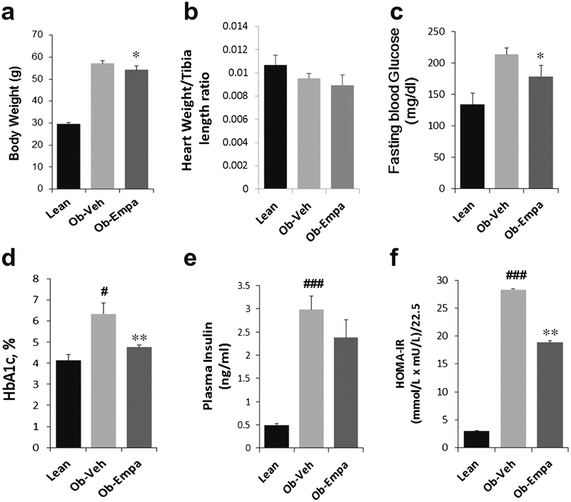
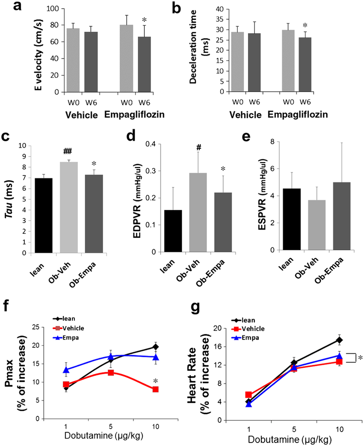
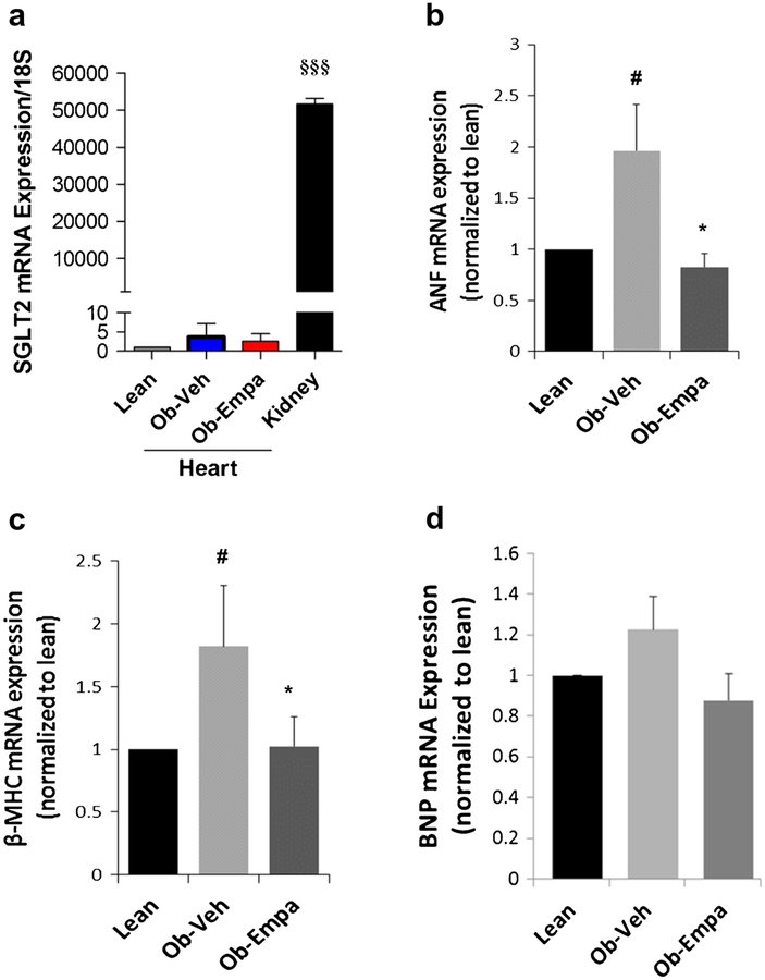
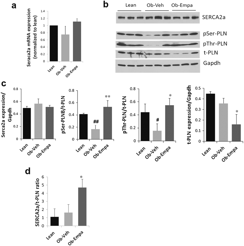
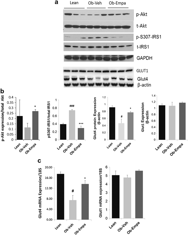
Methods and results: Cardiac phenotype was characterized by echocardiography, in vivo hemodynamics, histology, and molecular profiling. Our results demonstrate that empagliflozin significantly lowered HbA1c and slightly reduced body weight compared to vehicle treatment with no obvious changes in insulin levels. Empagliflozin also improved LV maximum pressure and in vivo indices of diastolic function. While systolic function was grossly not affected in both groups at steady state, response to dobutamine stimulation was significantly improved in the empagliflozin-treated group, suggesting amelioration of contractile reserve. This was paralleled by an increase in phospholamban (PLN) phosphorylation and increased SERCA2a/PLN ratio, indicative of enhanced SERCA2a function, further supporting improved cardiac relaxation and diastolic function. In addition, empagliflozin reconciled diabetes-associated increase in MAPKs and dysregulated phosphorylation of IRS1 and Akt, leading to improvement in myocardial insulin sensitivity and glucose utilization.

Conclusion: The data show that chronic treatment with empagliflozin improves diastolic function, preserves calcium handling and growth signaling pathways and attenuates myocardial insulin resistance in ob/ob mice, findings suggestive of a potential clinical utility for empagliflozin in the treatment of diastolic dysfunction.

Keywords: Calcium handling; Diabetes; Diastolic dysfunction; Empagliflozin; SGLT2 inhibitor; ob/ob mice.


Figures

Fig. 1
Fig. 1
General features of animals: a body weight, b heart weight/tibia length ratio, c fasting blood glucose, d HbA1c, e plasma insulin levels, and f HOMA-IR index of lean, ob/ob + vehicle (Ob-Veh) and ob/ob + empagliflozin (Ob-Empa). The data are expressed as mean ± s.d. (n = 10–15/group). *p = 0.05 and **p < 0.01 vs. Ob-Veh; #p < 0.05 and ###p < 0.001 vs. lean
Fig. 2
Fig. 2
Cardiac function measured by echocardiography and in vivo invasive hemodynamics. a E wave, mitral inflow peak velocity and b E wave deceleration time in ob/ob + vehicle and ob/ob + empagliflozin before (W0) and after (W6) treatment (n = 10/group);*p < 0.05 and *p < 0.05 vs. W0. c Tau and d EDPVR (end-diastolic pressure-volume relationship)—indexes of diastolic function. e ESPVR (end-systolic pressure volume relationship a measure of contractility; no significant effect but a trend to increase) in lean, ob/ob + vehicle (Ob-Veh) and ob/ob + empagliflozin (Ob-Empa) mice (n = 5–10/group); *p < 0.05 vs. Ob-Veh; #p < 0.05 vs. lean. e, f Dobutamine stimulation. Maximum pressure (Pmax, left) and heart rates (HR, right) of lean, ob/ob + vehicle and ob/ob + empagliflozin mice. Dobutamine stress effect is blunted in vehicle but responsive in empagliflozin group, while HR is significantly increased in empagliflozin group (n = 5–10). The % of increase was calculated by normalizing the dobutamine values to the steady state (i.e., nonstimulation) values. *p < 0.05 vs. Ob-Veh
Fig. 3
Fig. 3
SGLT2 cardiac expression cardiac hypertrophic markers. mRNA expression of SGLT2 (a), ANF (b), beta-MHC (c), and BNP (d) were evaluated by real-time quantitative PCR in heart samples from lean, ob/ob + vehicle (Ob-Veh) and ob/ob + empagliflozin (Ob-Empa). mRNAvalues were normalized to S18 RNA and plotted as a function of lean values. The data are expressed as mean ± s.d.(n = 5–7). §§§p < 0.0001 vs. Ob-Empa; *p < 0.05 vs. Ob-Veh;#p < 0.05 vs. lean
Fig. 4
Fig. 4
Expression of calcium cycling proteins. a mRNA expression of Serca2a. b representative western blots of SERCA2a and phospholamban (phospho-Serine-PLB—pSer-PLB, phospho-threonine PLB—pThr-PLB, and total t-PLB). c Densitometric quantification of corresponding cross-reactive bans in hearts samples from lean, ob/ob + vehicle (Ob-Veh) and ob/ob + empagliflozin (Ob-Empa). d SERCA2a/PLN ratio. mRNA values were normalized to S18 RNA and plotted as a function of lean values. Gapdh is used as a loading control. The data are expressed as mean ± s.d. *p < 0.05 and **p = 0.01 vs. Ob-Veh; #p < 0.05 and ##p = 0.01 vs. lean
Fig. 5
Fig. 5
Akt and IRS1 phosphorylation, Glut4 expression, and MAPKs activation. Western blot analyses of a Akt phosphorylation (p-Akt) and total Akt (t-Akt), serine phosphorylation of IRS1 (p-S307-IRS1) and total IRS1 (t-IRS1), Glut1 and Glut4 protein expression and d ERK, JNK, and p38 phosphorylation (p-ERK, p-JNK, p-p38) and total (t-ERK, t-JNK, t-p38) in hearts samples from lean, ob/ob + vehicle (Ob-Veh), and ob/ob + empagliflozin (Ob-Empa). b and e Respective densitometric measurements of phospho-immunoreactive bands normalized to total protein values for each protein are shown. Glut1 and Glut4 are normalized to β-actin. c mRNA expression of Glut4 and Glut1 in hearts samples from lean, ob/ob + vehicle (Ob-Veh) and ob/ob + empagliflozin (Ob-Empa). GAPDH and β-actin were used as loading control. The data are expressed as mean ± s.d. *p < 0.05, **p < 0.01 and ***p < 0.01 vs. Ob-Veh; #p < 0.05 and ###p < 0.001 vs. lean
Fig. 5
Fig. 5
Akt and IRS1 phosphorylation, Glut4 expression, and MAPKs activation. Western blot analyses of a Akt phosphorylation (p-Akt) and total Akt (t-Akt), serine phosphorylation of IRS1 (p-S307-IRS1) and total IRS1 (t-IRS1), Glut1 and Glut4 protein expression and d ERK, JNK, and p38 phosphorylation (p-ERK, p-JNK, p-p38) and total (t-ERK, t-JNK, t-p38) in hearts samples from lean, ob/ob + vehicle (Ob-Veh), and ob/ob + empagliflozin (Ob-Empa). b and e Respective densitometric measurements of phospho-immunoreactive bands normalized to total protein values for each protein are shown. Glut1 and Glut4 are normalized to β-actin. c mRNA expression of Glut4 and Glut1 in hearts samples from lean, ob/ob + vehicle (Ob-Veh) and ob/ob + empagliflozin (Ob-Empa). GAPDH and β-actin were used as loading control. The data are expressed as mean ± s.d. *p < 0.05, **p < 0.01 and ***p < 0.01 vs. Ob-Veh; #p < 0.05 and ###p < 0.001 vs. lean
Fig. 6
Fig. 6
Myocardial apoptosis. Representative Western blot analysis and densitometric quantification of Bcl2 and Bax expression in hearts samples from lean, ob/ob + vehicle (Ob-Veh) and ob/ob + empagliflozin (Ob-Empa). Data normalized to Gapdh expression. The data are expressed as mean ± s.d. #p < 0.05 vs. lean
Fig. 7
Fig. 7
Cardiac fibrosis by histology. a Representative 1.5×, 20×, and 40× micrographs of Trichrome-stained LV sections and b fibrosis quantification from lean, ob/ob + vehicle (Vehicle) and ob/ob + empagliflozin (Empagliflozin) (n = 5)

Comment in

References

    1. Abel ED, Litwin SE, Sweeney G. Cardiac remodeling in obesity. Physiol Rev. 2008;88:389–419. - PMC - PubMed
    1. Riva E, Andreoni G, Bianchi R, Latini R, Luvara G, Jeremic G, Traquandi C, Tuccinardi L. Changes in diastolic function and collagen content in normotensive and hypertensive rats with long-term streptozotocin-induced diabetes. Pharmacol Res. 1998;37:233–40. - PubMed
    1. Singleton JR, Smith AG, Russell JW, Feldman EL. Microvascular complications of impaired glucose tolerance. Diabetes. 2003;52: 2867–73. - PubMed
    1. Hemmingsen B, Lund SS, Gluud C, Vaag A, Almdal TP, Hemmingsen C, Wetterslev J. Targeting intensive glycaemic control versus targeting conventional glycaemic control for type 2 diabetes mellitus. The Cochrane Database of Systematic Reviews. 2013;11:Cd008143. - PubMed
    1. Bennett WL, Maruthur NM, Singh S, Segal JB, Wilson LM, Chatterjee R, Marinopoulos SS, Puhan MA, Ranasinghe P, Block L, et al. Comparative effectiveness and safety of medications for type 2 diabetes: an update including new drugs and 2-drug combinations. Ann Intern Med. 2011;154:602–13. - PMC - PubMed

MeSH terms