. 2017 Dec;3(1):25.
doi: 10.1186/s40729-017-0086-2. Epub 2017 Jun 22.

Ridge preservation using an in situ hardening biphasic calcium phosphate (β-TCP/HA) bone graft substitute-a clinical, radiological, and histological study

Affiliations

Ridge preservation using an in situ hardening biphasic calcium phosphate (β-TCP/HA) bone graft substitute-a clinical, radiological, and histological study

Ashish Kakar et al. Int J Implant Dent. 2017 Dec.

Abstract

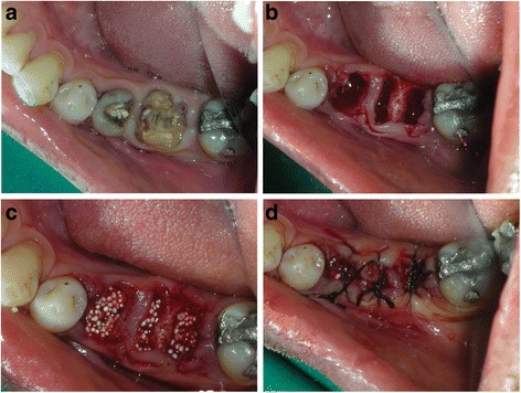
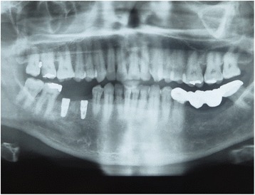
Background: Post-Extraction ridge preservation using bone graft substitutes is a conservative technique to maintain the width of the alveolar ridge. The objective of the present study was to evaluate an in situ hardening biphasic (HA/β-TCP) bone graft substitutes for ridge preservation without primary wound closure or a dental membrane.

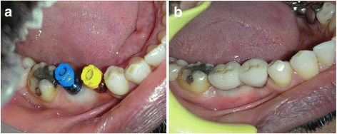
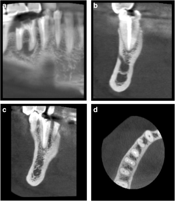
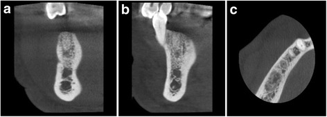
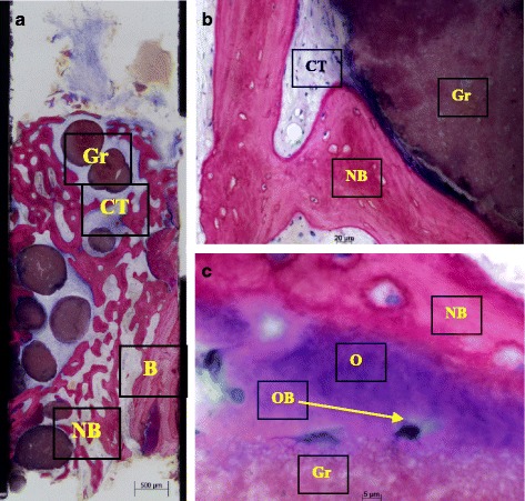
Methods: A total of 15 patients reported for tooth extraction were enrolled in this study. Implants were placed in average 5.2 ± 2 months after socket grafting. At this visit, Cone Beam CT (CBCT) images and core biopsies were taken. Implant stability (ISQ) was assessed at the insertion as well as at the day of final restoration.

Results: CBCT data revealed 0.79 ± 0.73 mm ridge width reduction from grafting to implant placement. Histomorphometric analysis of core biopsy samples revealed in average 21.34 ± 9.14% of new bone in the grafted sites. Primary implant stability was high (ISQ levels 70.3 ± 9.6) and further increased until final restoration.

Conclusions: The results of this study show that grafting of intact post-extraction sockets using a biphasic in situ hardening bone graft substitute results in an effective preservation of the ridge contour and sufficient new bone formation in the grafted sites, which is imperative for successful implant placement.


Figures

Fig. 1
Fig. 1
a Clinical occlusal view with fractured 45 and 46. b Post-extraction view of the socket. Note minimal trauma to the soft tissue and no flap reflection on the surgical site. c Graft material condensed into the extraction sockets showing good initial graft stability. d Black silk sutures placed with tissue approximation and no releasing incision in the flaps
Fig. 2
Fig. 2
a Clinical postoperative view after 4 months. Note that the healing was achieved only with tissue approximation. A good width of keratinized tissue is visible along with ridge preservation. Ready for implant placement in the grafted areas. b Implant placed in 45 area. Core biopsy sample taken from area 46. Note the integration of graft particles in the preserved alveolar ridge also inside the osteotomy site of 46. c Two Xive (Dentsply) implants placed in the preserved ridge. d. Postoperative X ray showing the implant positions in the mandible where the teeth were extracted and ridge preservation was accomplished
Fig. 3
Fig. 3
Postoperative X ray showing the implant positions in the mandible where the teeth were extracted and ridge preservation was accomplished
Fig. 4
Fig. 4
a Second stage surgery followed by impression making. Note the excellent width of keratinized tissue which was also preserved. b Implant crowns placed and loaded after 3 months of placement
Fig. 5
Fig. 5
CBCT images of the extraction site. a Preoperative CBCT showing fractured and un-restorable teeth #45 and #46 planned to be extracted. bd Cross sectional views
Fig. 6
Fig. 6
ac Four-month postoperative CBCT showing graft integration and preservation of ridge without collapse of the buccal or lingual cortical plates also showing the cross sections in the grafted area
Fig. 7
Fig. 7
ac Histological sections of bone core biopsy taken from the site of implantation using a trephine bur. a Overview image of coronal-apical cut through the entire core biopsy showing formation of new bone (NB) next to old bone of the extraction socket (B). easy-graft CRYSTAL particles (Gr) are embedded in well perfused connective tissue (CT) and new bone (NB) (Azur II and Pararosanilin, original magnification ×50). b Integration of easy-graft CRYSTAL particle (Gr) into newly formed bone (NB) and connective tissue (CT) showing tight contact between graft particle and new bone. c High magnification (×200) images of the interface between graft particle (Gr) and new bone (NB) showing osteoblasts (OB) forming osteoid (O) and formation of new bone (NB) on the surface of easy-graft CRYSTAL particles (Gr) (Azur II and Pararosanilin, original magnification ×200)

Similar articles

Cited by

References

    1. Araujo MG, Sukekava F, Wennstrom J, Lindhe J. Ridge alterations following implant placement in fresh extraction sockets: an experimental study in the dog. J Clin Periodontol. 2005;32:645–652. doi: 10.1111/j.1600-051X.2005.00726.x. - DOI - PubMed
    1. Van der Weijden F, Dell'Acqua F, Slot DE DE. Alveolar bone dimensional changes of post-extraction sockets in humans: a systematic review. J Clin Periodontol. 2009;36(12):1048–1058. doi: 10.1111/j.1600-051X.2009.01482.x. - DOI - PubMed
    1. Schropp L, Wenzel A, Kostopaulos L, Karring T. Bone healing changes and soft tissue contour changes following single-tooth: a clinical and radiographic 12-month prospective study. Int J Periodontol Restor Dent. 2003;23:313–323. - PubMed
    1. Fickl S, Zuhr O, Wachtel H, Stappert CF, Stein JM, Hürzeler MB. Dimensional changes of the alveolar ridge contour after different socket preservation techniques. J Clini Periodontol. 2008;35(10):906–913. doi: 10.1111/j.1600-051X.2008.01305.x. - DOI - PubMed
    1. Barone A, Orlando B, Cingano L, Marconcini S, Derchi G, Covani U. A randomized clinical trial to evaluate and compare implants placed in augmented versus non-augmented extraction sockets: 3-year results. J Periodontol. 2012;83(7):836–846. doi: 10.1902/jop.2011.110205. - DOI - PubMed

LinkOut - more resources