

Microscopy visualisation confirms multi-species biofilms are ubiquitous in diabetic foot ulcers
- PMID: 28643380
- PMCID: PMC7949972
- DOI: 10.1111/iwj.12777
Microscopy visualisation confirms multi-species biofilms are ubiquitous in diabetic foot ulcers
Abstract
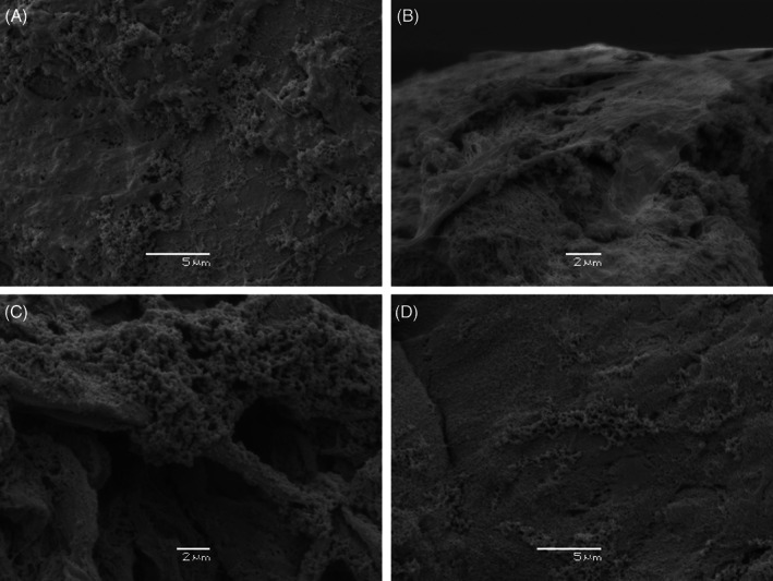
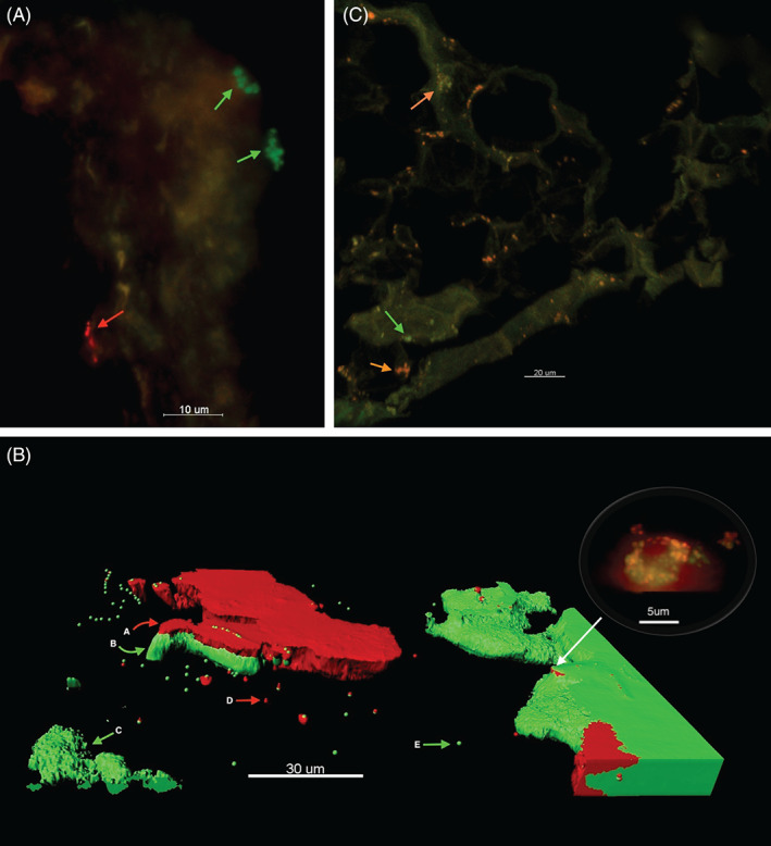
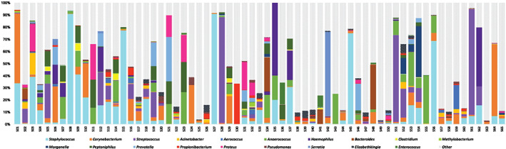
Increasing evidence within the literature has identified the presence of biofilms in chronic wounds and proposed that they contribute to delayed wound healing. This research aimed to investigate the presence of biofilm in diabetic foot ulcers (DFUs) using microscopy and molecular approaches and define if these are predominantly mono- or multi-species. Secondary objectives were to correlate wound observations against microscopy results in ascertaining if clinical cues are useful in detecting wound biofilm. DFU tissue specimens were obtained from 65 subjects. Scanning electron microscopy (SEM) and peptide nucleic acid fluorescent in situ hybridisation (PNA-FISH) techniques with confocal laser scanning microscopy (CLSM) were used to visualise biofilm structures. Next-generation DNA sequencing was performed to explore the microbial diversity. Clinical cues that included the presence of slough, excessive exudate, a gel material on the wound bed that reforms quickly following debridement, poor granulation and pyocyanin were correlated to microscopy results. Of the 65 DFU specimens evaluated by microscopy, all were characterised as containing biofilm (100%, P < 0·001). The presence of both mono-species and multi-species biofilms within the same tissue sections were detected, even when DNA sequencing analysis of DFU specimens revealed diverse polymicrobial communities. No clinical correlations were identified to aid clinicians in identifying wound biofilm. Microscopy visualisation, when combined with molecular approaches, confirms biofilms are ubiquitous in DFUs and form either mono- or multi-species biofilms. Clinical cues to aid clinicians in detecting wound biofilm are not accurate for use in DFUs. A paradigm shift of managing DFUs needs to consider anti-biofilm strategies.
Keywords: Biofilms; Diabetic foot ulcers; Fluorescent in situ hybridisation; Microscopy; Scanning electron microscopy.
© 2017 Medicalhelplines.com Inc and John Wiley & Sons Ltd.
Figures

References
-
- Lipsky BA, Berendt AR, Cornia PB, Pile JC, Peters EJ, Armstrong DG, Deery HG, Embil JM, Joseph WS, Karchmer AW, Pinzur MS, Senneville E; Infectious Diseases Society of America. 2012 Infectious Diseases Society of America clinical practice guideline for the diagnosis and treatment of diabetic foot infections. Clin Infect Dis 2012;54:e132–73. - PubMed
-
- Hurlow J, Bowler PG. Potential implications of biofilm in chronic wounds: a case series. J Wound Care 2012;21:109–19. - PubMed
-
- Lenselink E, Andriessen A. A cohort study on the efficacy of a polyhexanide‐containing biocellulose dressing in the treatment of biofilms in wounds. J Wound Care 2011;20:534–9. - PubMed
-
- Characklis WGMK. Biofilms. New York: John Wiley & Sons, 1990.
MeSH terms
Substances
LinkOut - more resources
Full Text Sources
Other Literature Sources
Medical
