. 2017 Dec;21(12):3394-3404.
doi: 10.1111/jcmm.13250. Epub 2017 Jun 23.

Knockdown of Mtfp1 can minimize doxorubicin cardiotoxicity by inhibiting Dnm1l-mediated mitochondrial fission

Affiliations

Knockdown of Mtfp1 can minimize doxorubicin cardiotoxicity by inhibiting Dnm1l-mediated mitochondrial fission

Lynn H H Aung et al. J Cell Mol Med. 2017 Dec.

Abstract

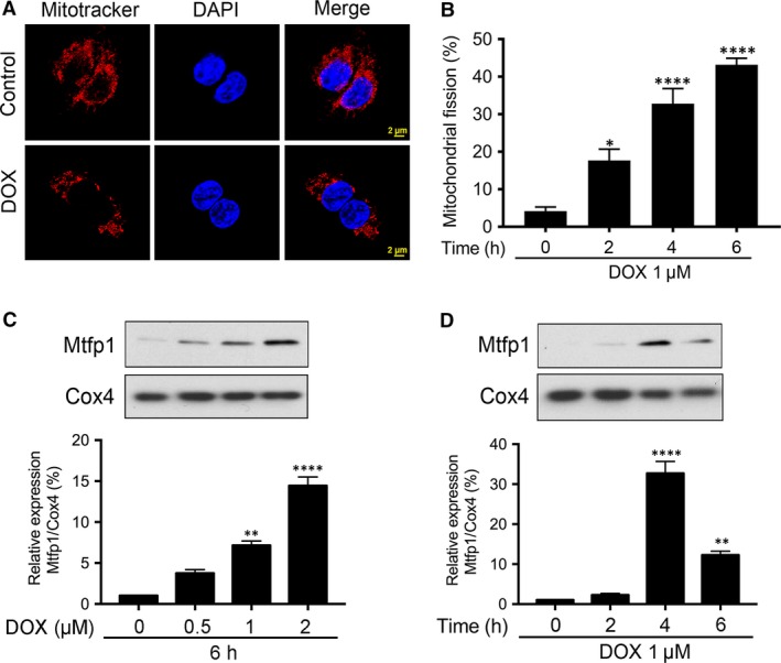
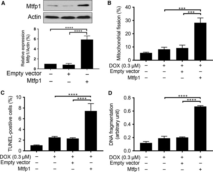
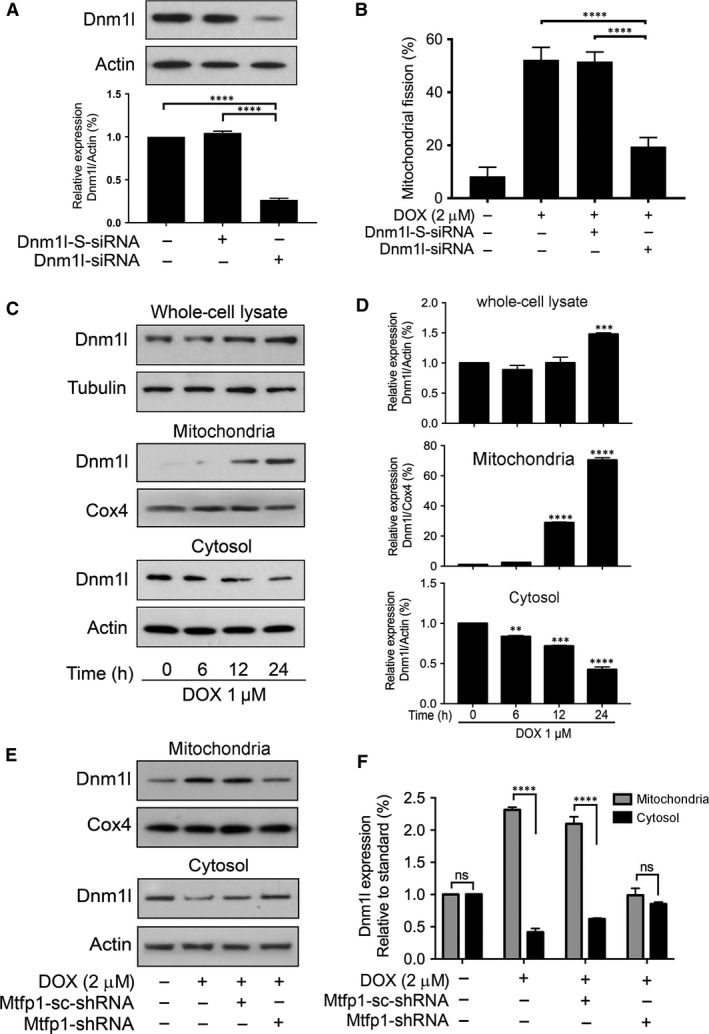
The long-term usage of doxorubicin (DOX) is largely limited due to the development of severe cardiomyopathy. Many studies indicate that DOX-induced cardiac injury is related to reactive oxygen species generation and ultimate activation of apoptosis. The role of novel mitochondrial fission protein 1 (Mtfp1) in DOX-induced cardiotoxicity remains elusive. Here, we report the pro-mitochondrial fission and pro-apoptotic roles of Mtfp1 in DOX-induced cardiotoxicity. DOX up-regulates the Mtfp1 expression in HL-1 cardiac myocytes. Knockdown of Mtfp1 prevents cardiac myocyte from undergoing mitochondrial fission, and subsequently reduces the DOX-induced apoptosis by preventing dynamin 1-like (Dnm1l) accumulation in mitochondria. In contrast, when Mtfp1 is overexpressed, a suboptimal dose of DOX can induce a significant percentage of cells to undergo mitochondrial fission and apoptosis. These data suggest that knocking down of Mtfp1 can minimize the cardiomyocytes loss in DOX-induced cardiotoxicity. Thus, the regulation of Mtfp1 expression could be a novel therapeutic approach in chemotherapy-induced cardiotoxicity.

Keywords: cardiotoxicity; doxorubicin; dyanmic-1-like (Dnm1l); mitochondrial fission; mitochondrial fission process 1 (Mtfp1).


Figures

Figure 1
Figure 1
Doxorubicin induces apoptosis in HL‐1 cardiac myocytes. A and B show caspase‐3 cleavage by immunoblot and densitometry; C and D show PARP1 cleavage by immunoblot and densitometry. Tubulin and β‐actin served as a loading control. (AD) Cells were stimulated with 1 μmol/l DOX and then harvested at the indicated time for immunoblotting. Figures presented are the representative figures of at least three independent experiments. The densitometry data were expressed as the mean ± SEM of three independent experiments. E shows DNA fragmentation. Cells were stimulated with 1 μmol/l DOX at indicated time‐points and apoptosis‐related DNA fragmentation were analysed using the cell death detection ELISA (E). Data were expressed as the mean ± SEM of three independent experiments. ns, non‐significant, *< 0.05, ***< 0.001, ****< 0.0001 versus 0 hr.
Figure 2
Figure 2
Doxorubicin‐induced mitochondrial fission is associated with up‐regulation in Mtfp1 expression. (A and B) doxorubicin (DOX) induces mitochondrial fission in HL‐1 cells. Cells were stimulated with 1 μmol/l DOX at indicated time‐points and mitochondrial morphology was analysed. A shows mitochondrial morphology. B shows percentage of cells undergoing mitochondrial fission. Data were expressed as the mean ± SEM of three independent experiments. (C and D) DOX up‐regulates mitochondrial fission process 1 (Mtfp1) expression in mitochondria in a dose‐ and time‐dependent manner. Analysis of Mtfp1 expression. HL‐1 cells were stimulated with the indicated doses of DOX and harvested at 6 hrs (C, upper panel), and cells were stimulated with 1 μmol/l DOX and then harvested at the indicated time (D, upper panel) for immunoblotting. Cytochrome‐c oxidase (Cox4) served as a loading control for mitochondrial fraction. Figures presented are the representative figures of at least three independent experiments. The densitometry data were expressed as the mean ± SEM of three independent experiments (C and D lower panels). *P < 0.05, **< 0.01, ****< 0.0001 versus non‐treatment.
Figure 3
Figure 3
Knockdown of Mtfp1 can prevent HL‐1 cell from doxorubicin‐induced mitochondrial fission and apoptosis. (A) Analysis of mitochondrial fission process 1 (Mtfp1) expression. Immunoblot shows Mtfp1 knockdown in HL‐1 cells. β‐actin served as a loading control. The densitometry data were expressed as the mean ± SEM of three independent experiments (lower panel). B shows mitochondrial morphology. Bar = 2 μm. C shows percentage of cells with mitochondrial fission. Cells were exposed to a higher concentration (2 μmol/l) doxorubicin (DOX) and mitochondrial fission was analysed by MitoTraker Red (B and C). D shows TUNEL‐positive cells. Bar = 20 μm. E shows percentage of TUNEL‐positive cells. F shows apoptosis‐related DNA fragmentation. Cells were exposed to a higher concentration (2 μmol/l) DOX, and apoptosis was analysed by TUNEL assay (D and E), and DNA fragments were analysed using the cell death detection ELISA (F). Figures presented are the representative figures of at least three independent experiments. Data were expressed as the mean ± SEM of three independent experiments. ***< 0.001 and ****< 0.0001.
Figure 4
Figure 4
Enforced expression of Mtfp1 sensitizes HL‐1 cells to doxorubicin‐induced mitochondrial fission and apoptosis. (A) Analysis of Mtfp1 expression. Immunoblot shows Mtfp1 overexpression in HL‐1 cells (upper panel). β‐actin served as a loading control. The densitometry data were expressed as the mean ± SEM of three independent experiments (lower panel). (B) Enforced expression of Mtfp1 sensitizes cells to undergo DOX‐induced mitochondrial fission. HL‐1 cells were exposed to a lower concentration of DOX. B shows percentage of cells with mitochondrial fission; (C and D) Enforced expression of Mtfp1 sensitizes cells to undergo DOX‐induced apoptosis. Cells were exposed to a lower concentration (0.3 μmol/l), and percentages of apoptosis were analysed by TUNEL assay (C) and DNA fragments were analysed using the cell death detection ELISA (D). Data were expressed as the mean ± SEM of three independent experiments. Figures presented are the representative figures of at least three independent experiments. ***< 0.001 and ****< 0.0001.
Figure 5
Figure 5
Dynamin 1‐like protein (Dnm1l) is predicted to be a Mtfp1's target protein. A shows schematic representation of the Mtfp1's target protein interaction network. B shows the confidence interaction scores of potential functionally associated proteins. Protein interaction network was constructed by STRING v10. Dnm1l shows highest estimated confidence score among all the potential functionally associated proteins.
Figure 6
Figure 6
Dnm1l is required for doxorubicin‐induced mitochondrial fission and Mtfp1 promotes doxorubicin‐induced Dnm1l accumulation in mitochondria. (A and B) Dnm1l is required for DOX to induce mitochondrial fission. (A) Analysis of Dnm1l expression. Immunoblot shows Dnm1l knockdown in HL‐1 cells. HL‐1 cells were transfected with scrambled siRNA, or Dnm1l siRNA, respectively, and after 48 hrs, were harvested for immunoblot (upper panel). Lower panel shows the densitometry. β‐actin served as a loading control. (B) Knockdown of Dnm1l inhibits doxorubicin (DOX)‐induced mitochondrial fission. B shows percentage of cells with mitochondrial fission. (C and D) DOX induces translocation of Dnm1l from cytosol to mitochondria. C shows Dnm1l expression by immunoblot. D shows the densitometry. **< 0.01, ***< 0.001 and ****< 0.0001 versus 0 hr. (E and F) Knockdown of Mtfp1 inhibits DOX‐induced Dnm1l accumulation in mitochondria. Then, they were treated with a higher concentration (2 μmol/l) of DOX for 24 hrs and harvested for subcellular fraction, and protein expression levels in different cellular compartments were analysed by immunoblot. E shows Dnm1l expression in subcellular fraction and F shows densitometry. The densitometry data were expressed as the mean ± SEM of three independent experiments (lower panel). β‐actin served as a loading control for whole‐cell lysate and cytosolic component. Cox4 served as a loading control for mitochondrial component. Figures presented are the representative figures of at least three independent experiments. Data were expressed as the mean ± SEM of three independent experiments. ns, non‐significant; ****< 0.0001.

References

    1. De Iuliis F, Salerno G, Corvino R, et al Anthracycline‐free neoadjuvant chemotherapy ensures higher rates of pathologic complete response in breast cancer. Clin Breast Cancer. 2017; 1: 34–40. - PubMed
    1. Karch A, Koch A, Grunwald V. A phase II trial comparing pazopanib with doxorubicin as first‐line treatment in elderly patients with metastatic or advanced soft tissue sarcoma (EPAZ): study protocol for a randomized controlled trial. Trials. 2016; 17: 312. - PMC - PubMed
    1. Khaliq NU, Sandra FC, Park DY, et al Doxorubicin/heparin composite nanoparticles for caspase‐activated prodrug chemotherapy. Biomaterials. 2016; 101: 131–42. - PubMed
    1. Singal PK, Iliskovic N. Doxorubicin‐induced cardiomyopathy. N Engl J Med. 1998; 339: 900–5. - PubMed
    1. Rosen GM, Halpern HJ. Spin trapping biologically generated free radicals: correlating formation with cellular injury. Methods Enzymol. 1990; 186: 611–21. - PubMed

MeSH terms