. 2017 Sep;246(9):670-681.
doi: 10.1002/dvdy.24537. Epub 2017 Jul 19.

IRF6 expression in basal epithelium partially rescues Irf6 knockout mice

Affiliations

IRF6 expression in basal epithelium partially rescues Irf6 knockout mice

Youssef A Kousa et al. Dev Dyn. 2017 Sep.

Abstract

Background: Mutations in IRF6, CHUK (IKKA), and RIPK4 can lead to a disease spectrum that includes cutaneous, limb, and craniofacial malformations. Loss of these alleles in the mouse leads to perinatal lethality and severe cutaneous, limb, and craniofacial defects also. Genetic rescue in the mouse has been shown for Ikka and Ripk4.

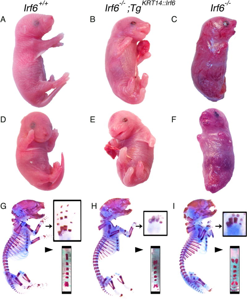
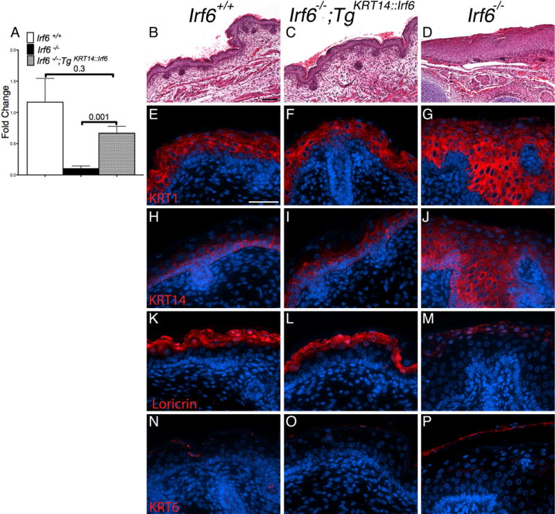
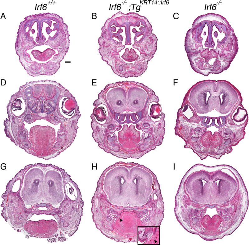
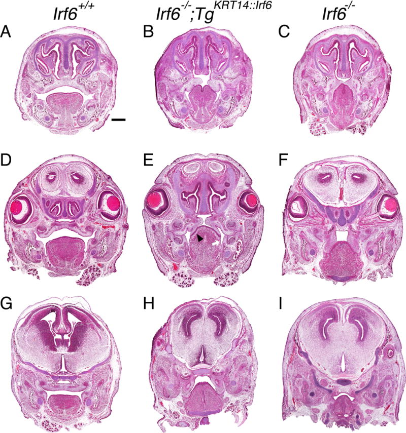
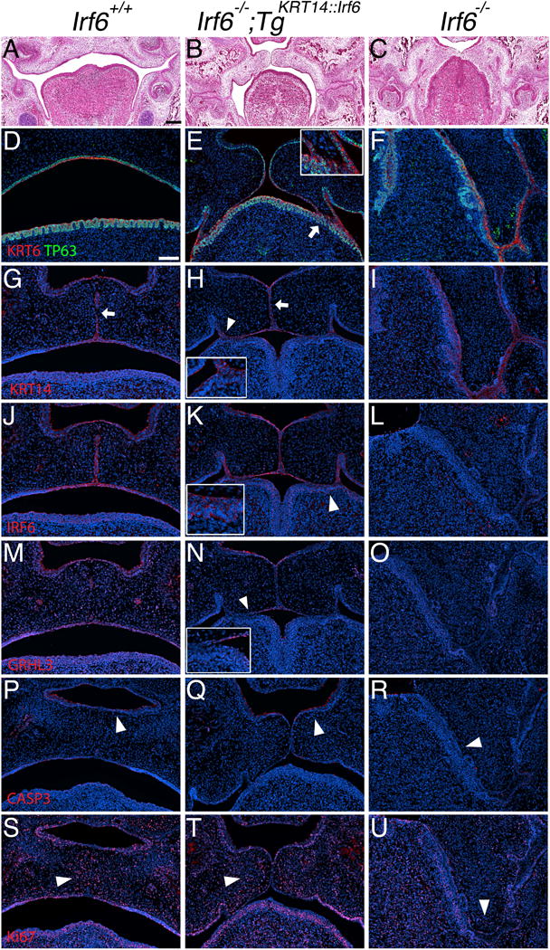
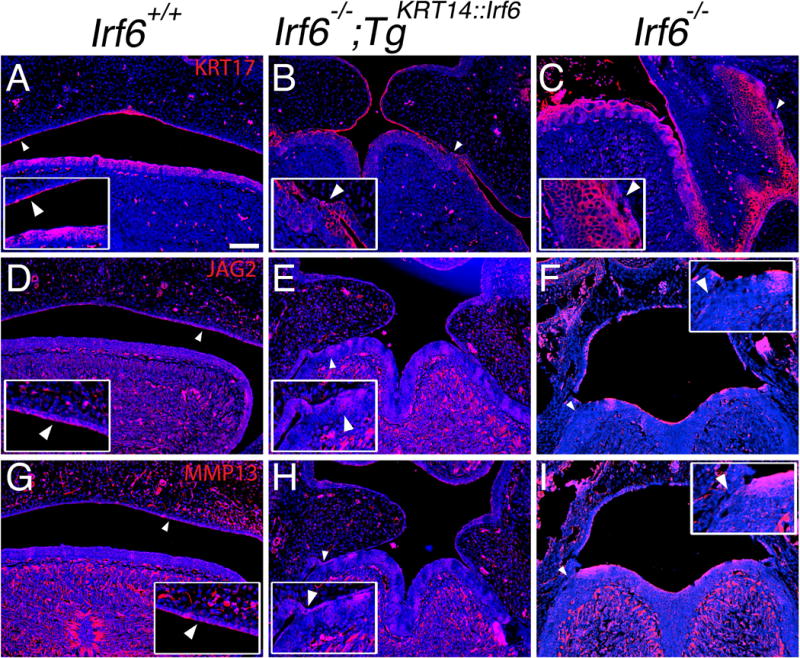
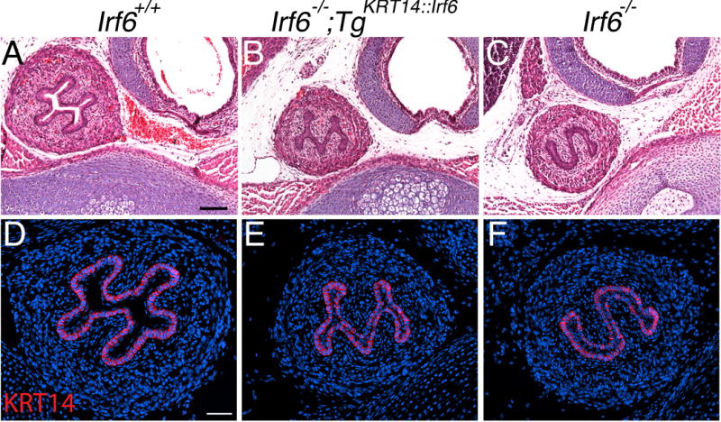
Results: Here, we show partial genetic rescue of Irf6 knockout embryos using the KRT14 promoter to drive Irf6 expression in the basal epithelium. In contrast to Irf6 knockout embryos, rescue embryos survive the immediate perinatal period. Macroscopic examination reveals rescue of skin adhesions between the axial and appendicular skeleton. Unexpectedly, KRT14-driven Irf6 expression does not completely rescue orofacial clefting and adhesions between the palate and tongue, suggesting the importance of cell-autonomous IRF6 expression in periderm. Like knockout embryos, Irf6 rescue embryos also have persistent esophageal adhesions, which likely contribute to postnatal demise.

Conclusions: Together, these data suggest that targeted expression of IRF6 can significantly reduce disease severity, but that a minimum level of Irf6 in both periderm and basal epithelial cells is necessary for orofacial development. Therefore, homologous human and mouse phenotypes are observed for IRF6, IKKA, and RIPK4. In this work, we show that altering the expression level of IRF6 dramatically modified this phenotype in utero. Developmental Dynamics 246:670-681, 2017. © 2017 Wiley Periodicals, Inc.

Keywords: GRHL3; IRF6; Van der Woude syndrome; cleft lip and palate; epidermis; epithelium; genetic rescue; limb defects; mouse models; oral periderm; popliteal pterygium syndrome.


Figures

Figure 1
Figure 1
Transgenic KRT14::Irf6 delays perinatal lethality but does not rescue limb skeletal defects. Profile (A–C and G–I) and Frontal (D–F) views of representative P0 pups with the following genotypes; (A, D, G) Irf6+/+left column; (B, E, H) Irf6−/−;TgKRT14::Irf6 center column; (C, F, I) Irf6−/− right column. Gross analysis of Irf6−/−;TgKRT14::Irf6 (rescue pups) reveals perinatal survival, limb clubbing and syndactyly, a curled tail and a somewhat taut, shiny skin compared to wildtype littermates. Unlike Irf6−/− (knockout) littermates, the appendages of rescue embryos are not attached to the body wall and an open oral cavity is visible. (G–I) Profile views of skeletal preparations of P0 pups. Analysis of rescue pups reveals limb clubbing and syndactyly, in a manner highly analogous to Irf6−/− (arrow, top inset). In contrast, the sternum (arrow head, bottom inset) and in particular the xiphoid process appears less bifurcated in rescue pups.
Figure 2
Figure 2
Irf6 expression using the KRT14 promoter rescues skin defects in knockout embryos. A) Analysis of skin mRNA levels reveals a significant increase in Irf6 expression in rescue (TgKRT14::Irf6) compared to knockout pups (Irf6−/−). No statistical differences are detected between rescue and wildtype pups (Irf6+/+). (B–D) Skin histological analysis (Hematoxylin and Eosin) reveals epidermal hypertrophy in (D) knockout but not in rescue (B) and wildtype (C) pups. E–P) Immunostaining of KRT1, KRT14, Loricrin and KRT6. (E–J) Rescue pups have epithelial stratification, without ectopic KRT1 and KRT14 expression. (K–M) Rescue pups demonstrate epithelial differentiation (Loricrin), similar to wild type embryos. (N–P) Irf6 knockout embryos have markers of cellular stress in mature skin (KRT6). Scale bars: (B–C) =100 μm; (E–P) = 50 μm.
Figure 3
Figure 3
Rescue embryos have oral adhesions and cleft palate at birth. Head coronal sections of perinatal pups stained with H&E. A–C) anterior, D–F) middle and G–I) posterior oral cavities of wildtype (A, D, G), rescue (B, E, H) and knockout (C, F, I) pups. The oral cavity of rescue pups is less severely affected overall but oral adhesions persist bilaterally in the anterior oral cavity (B) and in the middle oral cavity between the maxilla and mandible at the tooth germ (E). In the posterior palate of rescue embryos (H), adhesions and a fusion (black arrow head, enlarged with inset) is observed between the palate and the tongue. (A) Scale bar = 500 μm. T, Tongue; PS, palatal shelves.
Figure 4
Figure 4
Tongue-palate oral adhesions hinder palatal development in rescue embryos at E15.5. Head coronal sections stained with H&E in (A–C) anterior, (D–F) middle, (G–I) posterior sections of the oral cavity. Three comparison groups are illustrated, including (A, D, G) wildtype (Irf6+/+), (B, E, H) rescue (Irf6−/− ;TgKRT14::Irf6) and (C, F, I) knockout (Irf6−/−) embryos. Rescue embryos had a cleft palate in anterior (B), middle (E) and posterior (H) sections of the oral cavity. Oral adhesions between the mandible and maxilla were similar between rescue (E) and knockout embryos (F). In contrast, oral adhesions between the palate and the tongue were less severe in rescue embryos (E, black arrow head). We did not detect oral adhesions between palatal shelves and the mandible (E, white arrow). In posterior oral cavity, oral adhesions between the palatal shelves and tongue were less severe but the palatal shelves did not reorient toward midline or elevate. (A) Scale bar = 500 μm.
Figure 5
Figure 5
Oral adhesions between lingual and palatal epithelium prevents re-orientation and apposition of the palatal shelves. (A–U) Coronal section of E15.5 middle oral cavity with H&E (A–C) and immunostaining (D–U). Left column (images: A, D, G, J, M, P, S) shows wildtype embryos (Irf6+/+). Middle column (images: B, E, H, K, N, Q, T) shows rescue embryos (Irf6−/−;TgKRT14::Irf6). Right column (images: C, F, I, L, O, R, U) shows knockout embryos (Irf6−/−). (A) Wildtype embryos have a formed palate, whereas palatal shelves in (C) knockout embryos do not elevate. Palatal shelves in (B) rescue embryos (Irf6−/−;TgKRT14::Irf6) are able to re-orient and reach midline but do not fuse. (D–U) Immunostaining of (D–F) KRT6 (red) and TP63 (Green), (G–I) KRT14 (red), (J–L) IRF6 (red), (M–O) GRHL3 (red), (P–R) Activated Caspase 3 (red), (S–U) Ki- 67 (red). (D–F) In rescue embryos, TP63 marks the basal cell layer and KRT6 staining highlights the superficial epithelial layer. KRT6 marks periderm but was detected in areas of adhesion between the palate and the tongue (white arrow, enlarged with inset in top, right corner for rescue embryo) (E). (G–I) Unlike rescue embryos, loss of KRT14 immunostaining in wildtype embryos (G) indicates loss of the medial edge epithelium (white arrow). In rescue embryos, palatal shelves do not fuse even when there is apposition of the palatal shelves (H). Areas of epithelial adhesion show a KRT14 signal (white arrow head, enlarged with inset in bottom, left corner for rescue embryo). (J–L) Immunostaining shows a signal for IRF6 in the oral epithelium of rescue embryos (K), including sites of adhesion between the palate and tongue (white arrow head, enlarged with inset in bottom, left corner for rescue embryo). (M–O) In contrast to KRT6 expression in rescue embryos (compare E vs. N), we did not detect a GRHL3 signal in areas with palate-lingual oral adhesions (white arrow head, image N, enlarged with inset in bottom, left corner of rescue embryo). (P–R) A signal for Activated Caspase 3 was detected on the nasal surface of the palatal shelves in both (P) wildtype and (Q) rescue embryos but not in knockout littermates (white arrow head in each image). (S–U) Examination of proliferation shows a cluster of Ki-67-positive cells in the mesenchyme of (S) wildtype and (T) rescue embryos near the middle edge epithelium but not in (U) knockout littermates (white arrow head in each image). Scale bar (A) = 200 μm, (D) = 100 μm.
Figure 6
Figure 6
JAG2 and MMP13 expression in sites of oral adhesions between lingual and palatal epithelium. (AI) Coronal section of E15.5 middle oral cavity with immunostaining. Left column (images: A, D, G) shows wildtype embryos (Irf6+/+). Middle column (images: B, E, H) shows rescue embryos (Irf6−/− ;TgKRT14::Irf6). Right column (images: C, F, I) shows knockout embryos (Irf6−/−). Immunostaining of (AC) KRT17 (red), (D–F) JAG2 (red), (G–I) MMP13 (red), with nuclear staining (blue). (A–C) Compared to wildtype (A) and knockout (C) embryos, we found a discontinuous KRT17 expression pattern in rescue embryos (B) (white arrow head, enlarged with inset). (D–F) Unlike knockout embryos (F), we found increased JAG2 expression at sites of oral adhesions in rescue embryos (E) (white arrow head, enlarged with inset). (G–I) Similar to JAG2, MMP13 expression was increased at sites of oral adhesions in rescue embryos (H) compared to knockout embryos (I) (white arrow head, enlarged with inset in bottom, left corner for rescue embryo). Scale bar (A) =100 μm.
Figure 7
Figure 7
Rescue embryos have esophageal adhesions that contribute to delayed perinatal lethality. (A–C) H&E and (D–F) immunostaining staining of transverse sections from P0 pups of (A, D) wildtype (Irf6+/+), (B, E) rescue (Irf6−/−;TgKRT14::Irf6) and (C, F) knockout (Irf6−/−) sections. In stark contrast to the open lumen in wildtype pups (A, D), both rescue (B, E) and knockout (C, F) littermates have extensive esophageal adhesions. However, there was a distinct difference in the shape of the adhesion, with rescue pups having an “M” shaped adhesion while knockout embryos have had an “S” shaped adhesion. (D–F) Immunostaining for KRT14 revealed a signal but required a 10-fold greater fluorescent exposure time compared to skin. Scale bars (A) = 100 um, (B) = 50 um.

References

    1. BOTTI E, SPALLONE G, MORETTI F, MARINARI B, PINETTI V, GALANTI S, DE MEO PD, DE NICOLA F, GANCI F, CASTRIGNANO T, PESOLE G, CHIMENTI S, GUERRINI L, FANCIULLI M, BLANDINO G, KARIN M, COSTANZO A. Developmental factor IRF6 exhibits tumor suppressor activity in squamous cell carcinomas. Proc Natl Acad Sci U S A. 2011;108:13710–5. - PMC - PubMed
    1. BUSH JO, JIANG R. Palatogenesis: morphogenetic and molecular mechanisms of secondary palate development. Development. 2011;139:231–43. - PMC - PubMed
    1. DE LA GARZA G, SCHLEIFFARTH JR, DUNNWALD M, MANKAD A, WEIRATHER JL, BONDE G, BUTCHER S, MANSOUR TA, KOUSA YA, FUKAZAWA CF, HOUSTON DW, MANAK JR, SCHUTTE BC, WAGNER DS, CORNELL RA. Interferon regulatory factor 6 promotes differentiation of the periderm by activating expression of grainyhead-like 3. J Invest Dermatol. 2013;133:68–77. - PMC - PubMed
    1. FAKHOURI WD, RAHIMOV F, ATTANASIO C, KOUWENHOVEN EN, FERREIRA DE LIMA RL, FELIX TM, NITSCHKE L, HUVER D, BARRONS J, KOUSA YA, LESLIE E, PENNACCHIO LA, VAN BOKHOVEN H, VISEL A, ZHOU H, MURRAY JC, SCHUTTE BC. An etiologic regulatory mutation in IRF6 with loss- and gain-of-function effects. Hum Mol Genet. 2014;23:2711–20. - PMC - PubMed
    1. FAKHOURI WD, RHEA L, DU T, SWEEZER E, MORRISON H, FITZPATRICK D, YANG B, DUNNWALD M, SCHUTTE BC. MCS9.7 enhancer activity is highly, but not completely, associated with expression of Irf6 and p63. Dev Dyn. 2012;241:340–9. - PMC - PubMed

LinkOut - more resources