Review
. 2017 Dec;45(6):1901-1929.
doi: 10.1177/0300060516675504. Epub 2017 Jan 12.

Coronary microvascular dysfunction in diabetes mellitus

Affiliations
Review

Coronary microvascular dysfunction in diabetes mellitus

Aleksandar Kibel et al. J Int Med Res. 2017 Dec.

Abstract

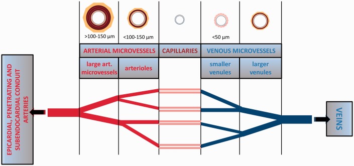
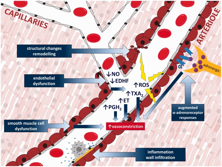
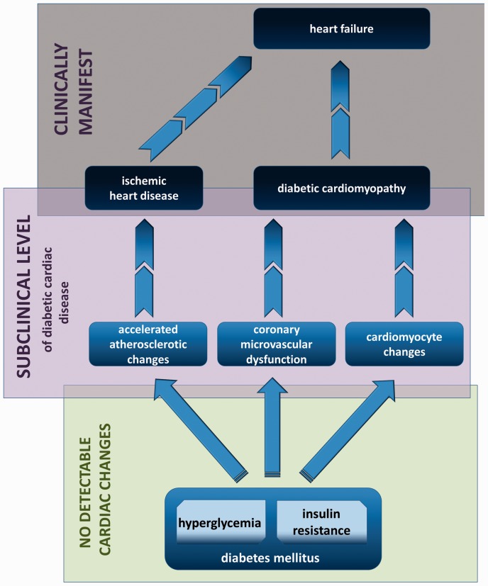
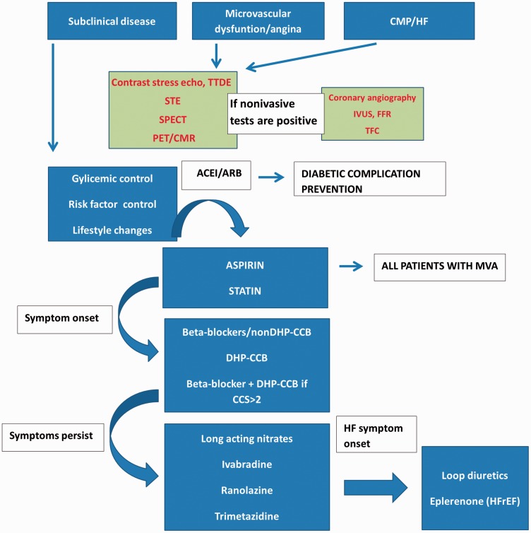
The significance, mechanisms and consequences of coronary microvascular dysfunction associated with diabetes mellitus are topics into which we have insufficient insight at this time. It is widely recognized that endothelial dysfunction that is caused by diabetes in various vascular beds contributes to a wide range of complications and exerts unfavorable effects on microcirculatory regulation. The coronary microcirculation is precisely regulated through a number of interconnected physiological processes with the purpose of matching local blood flow to myocardial metabolic demands. Dysregulation of this network might contribute to varying degrees of pathological consequences. This review discusses the most important findings regarding coronary microvascular dysfunction in diabetes from pre-clinical and clinical perspectives.

Keywords: Diabetes mellitus; arachidonic acid metabolites; artery; cardiomyopathy; coronary microvascular dysfunction; endothelial dysfunction; microcirculation; nitric oxide.


Figures

Figure 1.
Figure 1.
Overview of different microvessel categories in the heart. Arterial microvessels consist of a thin tunica intima, a thick tunica media composed of one to several layers of smooth muscle cells (“arterioles” possess 1–4 layers and are typically defined as <100–150 µm in diameter, whereas “large arterial microvessels” possess 4–6 layers, can be >100–150 µm in diameter and can also be called “small arteries”) and a tunica adventitia. Venous microvessels have thinner vascular walls compared with those of arterial microvessels, with venules <50 µm in diameter and not possessing smooth muscle cell layers. Smaller venules possess only endothelial cells and pericytes. From epicardial conduit arteries, numerous arterial branches arise that contribute to perfusion of the outer two-thirds of the myocardium. Penetrating arteries pass through the outer myocardial layers without branching. Ultimately, in the inner one-third of the myocardium, small branches arise from penetrating arteries to form the arterial plexus that perfuses the subendocardium.
Figure 2.
Figure 2.
Primary pathophysiologic mechanisms of coronary microvascular dysfunction. EDHF: endothelium-derived hyperpolarizing factor; ROS: reactive oxygen species; ET: endothelin; PGH2: prostaglandin H2; TXA2: thromboxane A2.
Figure 3.
Figure 3.
Clinical progression of diabetes-induced adverse cardiac changes.
Figure 4.
Figure 4.
Proposed diagnostic-therapeutic algorithm. There is no consensus or specific clinical guidelines for diabetic coronary microvascular dysfunction, because studies are limited and known information is scarce. Therapeutic strategies can, at this point, be based on preventive measures, treatment of diabetes (although intensive glycaemic control has not been proven to improve coronary microvascular dysfunction, it is important for preventing diabetic complications) and treatment of microvascular angina and/or cardiomyopathy/heart failure. Early detection/diagnosis and monitoring of disease progression are advisable and can contribute to timely initiation of therapeutic interventions. TTDE: transthoracic Doppler echocardiography; STE: SPECT-single photon emission tomography; PET: positron emission tomography; CMR: cardiac magnetic resonance; IVUS: intravascular ultrasound; FFR: fractional flow reserve; TFC: TIMI frame count; MVA: microvascular angina; BB: beta-blockers; CCB: calcium channel blockers; ACEI: angiotensin-converting enzyme inhibitor; ARB: angiotensin receptor blocker; CCS: Canadian Cardiac Society; CMP: cardiomyopathy; HF: heart failure; HFREF: heart failure with reduced ejection fraction.

References

    1. Leon BM, Maddox TM. Diabetes and cardiovascular disease: Epidemiology, biological mechanisms, treatment recommendations and future research. World J Diabetes 2015; 6: 1246–1258. - PMC - PubMed
    1. Kibel A. Could angiotensin-(1–7) be connected with improvement of microvascular function in diabetic patients? Angiotensin-(1–7) iontophoresis may provide the answer. Med Hypotheses 2016; 93: 16–20. 10.1016/j.mehy.2016.05.010. - DOI - PubMed
    1. Picchi A, Capobianco S, Qiu T, et al. Coronary microvascular dysfunction in diabetes mellitus: a review. World J Cardiol 2010; 2: 377–390. - PMC - PubMed
    1. Tabit CE, Chung WB, Hamburg NM, et al. Endothelial dysfunction in diabetes mellitus: molecular mechanisms and clinical implications. Rev Endocr Metab Disord 2010; 11: 61–74. - PMC - PubMed
    1. Tousoulis D, Papageorgiou N, Androulakis E, et al. Diabetes mellitus-associated vascular impairment: novel circulating biomarkers and therapeutic approaches. J Am Coll Cardiol 2013; 62: 667–676. - PubMed

LinkOut - more resources