

Advanced neuroimaging of carbon monoxide poisoning
- PMID: 28643616
- PMCID: PMC5602327
- DOI: 10.1177/1971400916689342
Advanced neuroimaging of carbon monoxide poisoning
Abstract
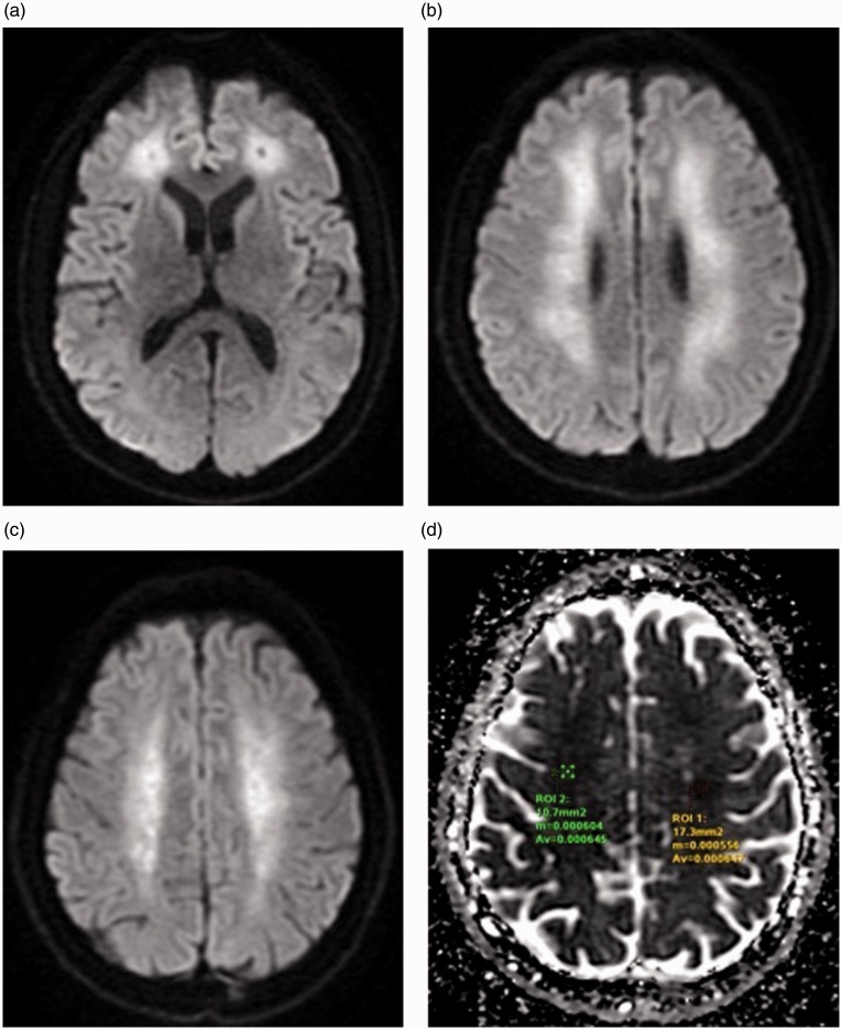
Carbon monoxide (CO) inhalation is nowadays the most common cause of fatal poisoning worldwide. CO binds to haemoglobin 230-270 times more avidly than oxygen, thus leading to formation of carboxyhaemoglobin with subsequent reduction of tissue oxygenation. Brain is mainly affected due to its high oxygen requirement. Up to two-thirds of patients who survive the acute phase of this pathology present a delayed leukoencephalopathy, usually in a period ranging from two to 40 days. White matter damage closely relates to long-term prognosis of these patients. On the other hand CO seems to play a fundamental role as a possible neuro-protective agent in ischaemic stroke. Diagnostic imaging, with computed tomography and magnetic resonance imaging, especially magnetic resonance spectroscopy, is very useful to depict the presence and extension of this pathology, in both acute and late phase. Nevertheless, a correlation of imaging studies with clinical history and laboratory data is fundamental to perform the correct diagnosis. The purpose of this article is to highlight the imaging features of brain CO poisoning in acute and late phase, describing a case report of a 56-year-old man found unconscious at home.
Keywords: Brain CO poisoning; diffusion magnetic resonance imaging; leukoencephalopathy; magnetic resonance spectroscopy.
Figures
References
-
- Bleecker ML. Carbon monoxide intoxication. Handb Clin Neurol 2015; 131: 191–203. - PubMed
-
- Park EJ, Min YG, Kim GW, et al. Pathophysiology of brain injuries in acute carbon monoxide poisoning: A novel hypothesis. Med Hypotheses 2014; 83: 186–189. - PubMed
-
- Chiew AL, Buckley NA. Carbon monoxide poisoning in the 21st century. Crit Care 2014; 18: 221.
-
- Hopkins RO, Woon FL. Neuroimaging, cognitive, and neurobehavioural outcomes following carbon monoxide poisoning. Behav Cogn Neurosci Rev 2006; 5: 141–155. - PubMed
Publication types
MeSH terms
LinkOut - more resources
Full Text Sources
Other Literature Sources
Medical
