Review
. 2017 Oct;52(5):503-542.
doi: 10.1080/10409238.2017.1337705. Epub 2017 Jun 23.

Lytic transglycosylases: concinnity in concision of the bacterial cell wall

Affiliations
Review

Lytic transglycosylases: concinnity in concision of the bacterial cell wall

David A Dik et al. Crit Rev Biochem Mol Biol. 2017 Oct.

Abstract

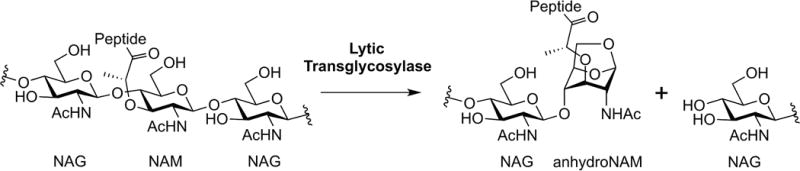
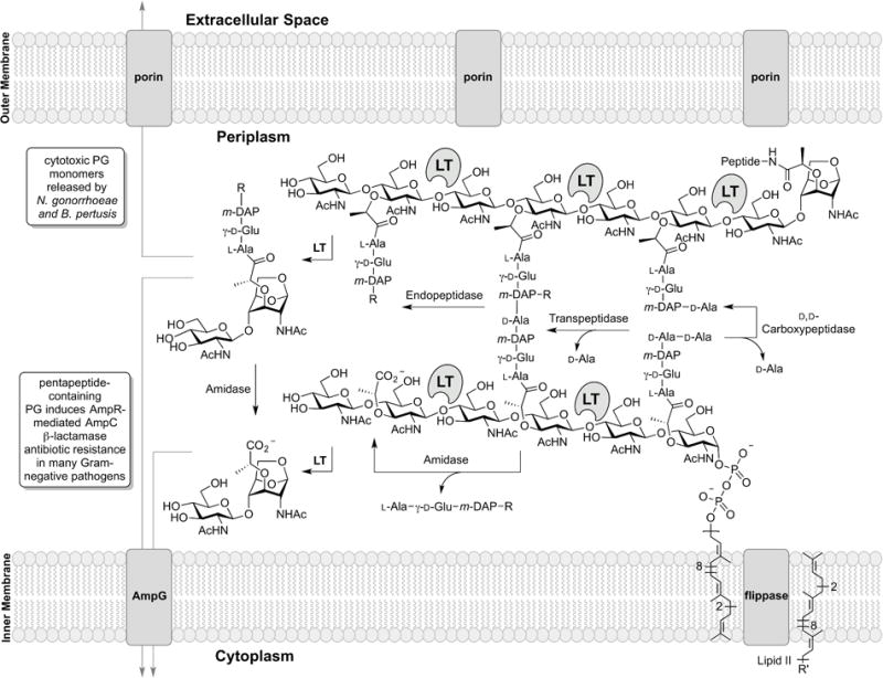
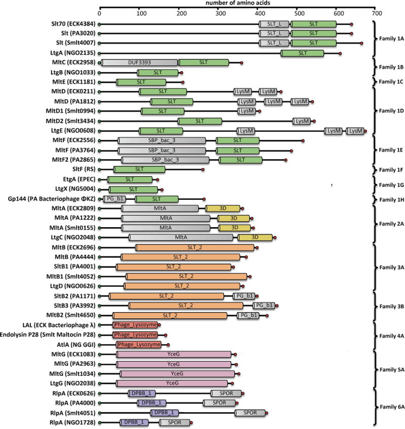
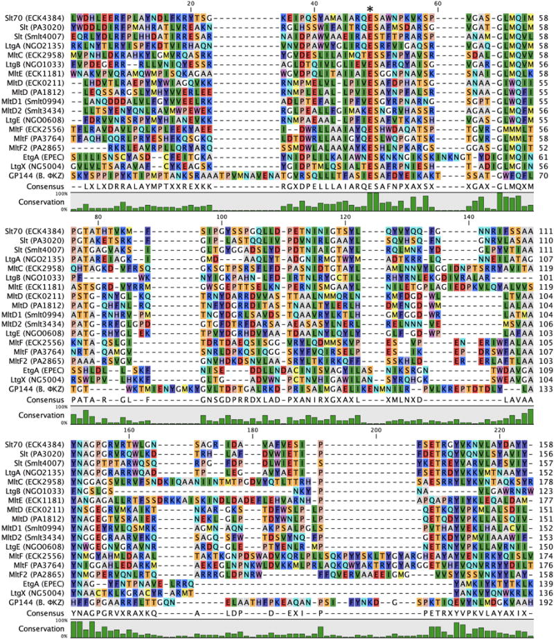
The lytic transglycosylases (LTs) are bacterial enzymes that catalyze the non-hydrolytic cleavage of the peptidoglycan structures of the bacterial cell wall. They are not catalysts of glycan synthesis as might be surmised from their name. Notwithstanding the seemingly mundane reaction catalyzed by the LTs, their lytic reactions serve bacteria for a series of astonishingly diverse purposes. These purposes include cell-wall synthesis, remodeling, and degradation; for the detection of cell-wall-acting antibiotics; for the expression of the mechanism of cell-wall-acting antibiotics; for the insertion of secretion systems and flagellar assemblies into the cell wall; as a virulence mechanism during infection by certain Gram-negative bacteria; and in the sporulation and germination of Gram-positive spores. Significant advances in the mechanistic understanding of each of these processes have coincided with the successive discovery of new LTs structures. In this review, we provide a systematic perspective on what is known on the structure-function correlations for the LTs, while simultaneously identifying numerous opportunities for the future study of these enigmatic enzymes.

Keywords: AmpC; AmpR; Lytic transglycosylase; bacteria; cell-wall recycling; muropeptide; peptidoglycan; secretion system.


Conflict of interest statement

Declaration of Interest:

The authors report no conflicts of interest. The authors alone are responsible for the content and writing of this article. This work was supported by a grant to S.M. from the National Institute of Health (NIH Grant GM61629). D.A.D. is a Fellow of the Chemistry-Biochemistry-Biology Interface (CBBI) Program (NIH Training Grant T32GM075762) and a Fellow of the ECK Institute of Global Health at the University of Notre Dame.

Figures

Figure 1
Figure 1
The hallmark reaction of the lytic transglycosylases.
Figure 2
Figure 2
The periplasmic events (where the LTs exist) of Gram-negative cell-wall recycling are depicted. The membrane anchoring of the LTs is not represented in the figure. The LT reaction sites are shown. In the scheme R = D-Ala-D-Ala (penta), or D-Ala (tetra), or H (tri) and R’ = diphosphoryl-N-acetyl muramoyl (glucosamine)-L-Ala-γ-D-Glu-meso-DAP-D-Ala-D-Ala.
Figure 3
Figure 3
Domain architecture of Gram-negative LTs. Domains were assigned based on consensus analysis of Pfam Database and NCBI conserved domain database. Protein name and locus tag are given for E. coli K12 (ECK…), P. aeruginosa PAO1 (PA…), S. maltophilia KJ (Smlt…), and N. gonorrhoeae FA1090 (NGO…). Locus tags are not given for Enteropathogenic E. coli (EPEC) or Rhodobacter sphaeroides (RS). For the bacteriophage LTs (family 1H and 4), the name of the infected host organism (ECK, PA, Smlt, and NG) and the identities of the bacteriophages encoding these LTs are provided. The PG_binding_1 domain of Family 1H and 3B is abbreviated as PG_b1. A color version of this figure is available at www.tandfonline.com/ibmg.
Figure 4
Figure 4
Multiple-sequence alignment displaying the LT domains of Gram-negative Family 1 LTs. In consideration of the size of the Family 1 alignment, R. sphaeroides SltF is not included as the SLT domain of the protein is considerably larger than the other Family 1 members. The asterisk denotes the position of the catalytic residue. A color version of this figure is available at www.tandfonline.com/ibmg.
Figure 5
Figure 5
X-ray structure alignment of Family 1 LTs, displaying the conservation of the core SLT domain (Pfam: PF01464) and the diversity of the peripheral domains. The ribbon representation of each apo LT crystal structure is displayed below with a transparent surface representation. The structure of an LT domain of Family 1D has not been solved, therefore only the peripheral domain is displayed. Family 1E is displayed in both the active and inactive conformations. Refer to table 1 for PDB codes. A color version of this figure is available at www.tandfonline.com/ibmg.
Figure 6
Figure 6
Multiple-sequence alignment displaying the LT domain of Gram-negative Family 2 LTs. The asterisk denotes the position of the catalytic residue. A color version of this figure is available at www.tandfonline.com/ibmg.
Figure 7
Figure 7
X-ray structure alignment of Gram-negative Family 2 LTs, displaying conservation of the 3D domain (Pfam: PF06725). The 3D domain is comprised of two subdomains linked by a hinge. The two structures show an open (E. coli MltA) and a closed (N. gonorrhoeae LtgC) protein conformation. The structure overlay aligns the top sub-domain. The bottom sub-domains are also conserved, but align differently in the open and closed states. The ribbon representation of each apo LT crystal structure is displayed to the right with a transparent surface representation. A color version of this figure is available at www.tandfonline.com/ibmg.
Figure 8
Figure 8
Multiple-sequence alignment displaying the LT domain of Gram-negative Family 3 LTs. The asterisk denotes the position of the catalytic residue. A color version of this figure is available at www.tandfonline.com/ibmg.
Figure 9
Figure 9
X-ray structure alignment of Gram-negative Family 3 LTs, displaying the conservation of the Slt_2 domain (Pfam: PF13406). The ribbon representation of each apo LT crystal structure is displayed below with a transparent surface representation. A color version of this figure is available at www.tandfonline.com/ibmg.
Figure 10
Figure 10
Multiple-sequence alignment displaying the LT domain of Gram-negative Family 4 LTs. The asterisk denotes the position of the catalytic residue. A color version of this figure is available at www.tandfonline.com/ibmg.
Figure 11
Figure 11
X-ray structure of the Gram-negative Family 4 LT bacteriophage endolysin λ; the Phage_Lysozyme domain (Pfam: PF00959) comprises most of the solved structure. The ribbon representation of the apo LT crystal structure is displayed with a transparent surface representation. A color version of this figure is available at www.tandfonline.com/ibmg.
Figure 12
Figure 12
Multiple-sequence alignment displaying the LT domain of Gram-negative Family 5 LTs. The asterisk denotes the position of the catalytic residue. A color version of this figure is available at www.tandfonline.com/ibmg.
Figure 13
Figure 13
X-ray structure alignment of Gram-negative Family 5 and Gram-positive Family 1 LTs, displaying the similar folds of the YceG domain (Pfam: PF02618) of Gram-negative Family 5 and the PG_Hydrolase_2 domain (Pfam: PF07486) of Gram-positive Family 1. Notably, the amino-acid sequence between the two domains is not similar. The ribbon representation of each apo LT crystal structure is displayed with a transparent surface representation. A color version of this figure is available at www.tandfonline.com/ibmg.
Figure 14
Figure 14
Multiple-sequence alignment displaying the LT domain of Gram-negative Family 6 LTs. The asterisk denotes the position of the catalytic residue. A color version of this figure is available at www.tandfonline.com/ibmg.
Figure 15
Figure 15
Domain architecture of Gram-positive LTs. Domains were assigned based on consensus analysis of Pfam Database and NCBI conserved-domain database. Protein names and those of the organisms are given for B. anthracis (BA), B. cereus (BC) and C. difficile (CD). Locus tags are not given. Notably, it is not known if the SleB homologue of C. difficile, which lacks the N-terminal PG_binding_1 (PG_b1) domain, is an LT, therefore it is not included. A color version of this figure is available at www.tandfonline.com/ibmg.
Figure 16
Figure 16
Multiple-sequence alignment displaying the LT domain of Gram-positive Family 1 LTs. The sequence of a C. difficile SleB homologue is included for comparison, although it is not known whether this protein is an LT. The asterisk denotes the position of the catalytic residue. A color version of this figure is available at www.tandfonline.com/ibmg.
Figure 17
Figure 17
Multiple-sequence alignment displaying the LT domain of Gram-positive Family 2 LTs. The asterisk denotes the position of the catalytic residue. A color version of this figure is available at www.tandfonline.com/ibmg.
Figure 18
Figure 18
X-ray structure alignment of Gram-positive Family 2 LTs, displaying the conservation of the SpoIID domain (Pfam: PF08486). The ribbon representation of each apo LT crystal structure is displayed below with a transparent surface representation. A color version of this figure is available at www.tandfonline.com/ibmg.
Figure 19
Figure 19
Proposed LT reaction mechanisms.
Figure 20
Figure 20
LT inhibitors: (A) the natural products bulgecin A-C, (B) NAG-thiazoline, (C) iminosaccharides, and (D) iminocyclitols.

References

    1. Abanes-De Mello A, Sun YL, Aung S, et al. A cytoskeleton-like role for the bacterial cell wall during engulfment of the Bacillus subtilis forespore. Genes Dev. 2002;16:3253–3264. - PMC - PubMed
    1. Adin DM, Engle JT, Goldman WE, et al. Mutations in ampG and lytic transglycosylase genes affect the net release of peptidoglycan monomers from Vibrio fischeri. J Bacteriol. 2009;191:2012–2022. - PMC - PubMed
    1. Adu-Bobie J, Lupetti P, Brunelli B, et al. GNA33 of Neisseria meningitidis is a lipoprotein required for cell separation, membrane architecture, and virulence. Infect Immun. 2004;72:1914–1919. - PMC - PubMed
    1. Alcorlo M, Martínez-Caballero S, Molina R, et al. Carbohydrate recognition and lysis by bacterial peptidoglycan hydrolases. Curr Opin Struct Biol. 2017;44:87–100. - PubMed
    1. Alvarez L, Hernandez SB, de Pedro MA, et al. Ultra-sensitive, high-resolution liquid chromatography methods for the high-throughput quantitative analysis of bacterial cell wall chemistry and structure. Methods Mol Biol. 2016;1440:11–27. - PubMed

Publication types