Observational Study
. 2018 Jan;38 Suppl 1(Suppl 1):S79-S88.
doi: 10.1097/IAE.0000000000001728.

ABNORMAL RETINAL REFLECTIVITY TO SHORT-WAVELENGTH LIGHT IN TYPE 2 IDIOPATHIC MACULAR TELANGIECTASIA

Affiliations
Observational Study

ABNORMAL RETINAL REFLECTIVITY TO SHORT-WAVELENGTH LIGHT IN TYPE 2 IDIOPATHIC MACULAR TELANGIECTASIA

Ferenc B Sallo et al. Retina. 2018 Jan.

Abstract

Purpose: Macular telangiectasia Type 2 (MacTel) is a bilateral, progressive, potentially blinding retinal disease characterized by vascular and neurodegenerative signs, including an increased parafoveal reflectivity to blue light. Our aim was to investigate the relationship of this sign with other signs of macular telangiectasia Type 2 in multiple imaging modalities.

Methods: Participants were selected from the MacTel Type 2 study, based on a confirmed diagnosis and the availability of images. The extent of signs in blue-light reflectance, fluorescein angiographic, optical coherence tomographic, and single- and dual-wavelength autofluorescence images were analyzed.

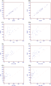
Results: A well-defined abnormality of the perifovea is demonstrated by dual-wavelength autofluorescence and blue-light reflectance in early disease. The agreement in area size of the abnormalities in dual-wavelength autofluorescence and in blue-light reflectance images was excellent: for right eyes: ρ = 0.917 (P < 0.0001, 95% confidence interval 0.855-0.954, n = 46) and for left eyes: ρ = 0.952 (P < 0.0001, 95% confidence interval 0.916-0.973, n = 49). Other changes are less extensive initially and expand later to occupy that area and do not extend beyond it.

Conclusion: Our findings indicate that abnormal metabolic handling of luteal pigment and physical changes giving rise to increased reflectance are widespread in the macula throughout the natural history of the disease, precede other changes, and are relevant to early diagnosis.


Conflict of interest statement

Proprietary interest: no conflicting relationship exists for any author.

Figures

Figure 1
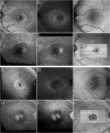
Figure 1. Aligned multimodal images of a MacTel eye with very early disease
A: 488nm (blue) reflectance image, B: dual-wavelength autofluorescence image (DWAF), C: single wavelength (488nm) fundus autofluorescence image, D: infrared image, E and F: early and late phase fluorescein angiograms, G: IR image with the en face view of the Ellipsoid Zone superimposed, H: IR image with the en face map of the External Limiting Membrane (ELM) superimposed. An eye with very early mactel disease from an asymmetrical case, demonstrating essentially no sign of mactel and only a few diabetes-related microaneurysms in the FFA. The FAF, DWAF images appear essentially normal, demonstrating the peak of luteal pigment density at the foveal center, that decreases towards the periphery. The dark central area in the EZ map corresponds to the rod-free center of the fovea externa with very high packing density of cones, where the EZ signal is normally weaker. The fellow eye (not shown) demonstrates all typical signs of MacTel.
Figure 2
Figure 2. BLR imaging in MacTel and multimodal correlates
Aligned multimodal images of a relatively early case (top panel) and a later-stage MacTel eye, bottom panel. A: blue light reflectance images, B: dual wavelength (488+514nm) autofluorescence images, C: single (488nm, blue) autofluorescence images, D: early phase FFA images, E: late-phase FFA images, F: topographic maps of the EZ (IS/OS layer). The small, round, hyper-reflective area close to the fovea in the BLR image in the top panel is an artifact due to internal reflections within the lens system of the Spectralis SLO system.
Figure 3
Figure 3. Correlation of lesion extent in multimodal images of type 2 MacTel
Top row: BLR vs DWAF area in (A) right and (B) in left eyes. Second row: DWAF area vs single wavelength blue light AF area, in right (C) and in left eyes (D). NB: in 28/96 eyes the AF lesion was not possible to delineate due to an indistinct peripheral boundary. Third row: BLR hyper-reflective area vs late FFA area, in right (E) and in left eyes (F). Bottom row: BLR hyper-reflective area vs EZ (IS/OS) break area, in right (G) and in left eyes (H). All data shown are expressed in ×1000 pixels (proprietary units).

References

    1. Gass JD, Oyakawa RT. Idiopathic juxtafoveolar retinal telangiectasis. Arch Ophthalmol. 1982;100:769–80. - PubMed
    1. Charbel Issa P, Gillies MC, Chew EY, et al. Macular telangiectasia type 2. Prog Retin Eye Res. 2013;34:49–77. - PMC - PubMed
    1. Powner MB, Gillies MC, Tretiach M, et al. Perifoveal muller cell depletion in a case of macular telangiectasia type 2. Ophthalmology. 2010;117:2407–16. - PMC - PubMed
    1. Powner MB, Gillies MC, Zhu M, et al. Loss of Muller's cells and photoreceptors in macular telangiectasia type 2. Ophthalmology. 2013;120:2344–52. - PubMed
    1. Clemons TE, Gillies MC, Chew EY, et al. Baseline characteristics of participants in the natural history study of macular telangiectasia (MacTel) MacTel Project Report No. 2. Ophthalmic Epidemiol. 17:66–73. - PMC - PubMed

Publication types

MeSH terms

Supplementary concepts