. 2017 Jul;9(7):698-707.
doi: 10.1038/nchem.2714. Epub 2017 Feb 6.

Post-translational modification of ribosomally synthesized peptides by a radical SAM epimerase in Bacillus subtilis

Affiliations

Post-translational modification of ribosomally synthesized peptides by a radical SAM epimerase in Bacillus subtilis

Alhosna Benjdia et al. Nat Chem. 2017 Jul.

Abstract

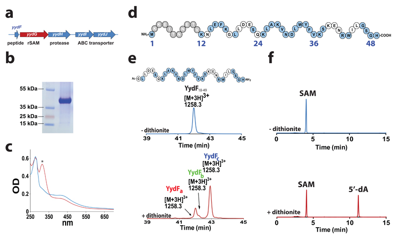
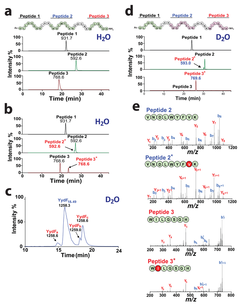
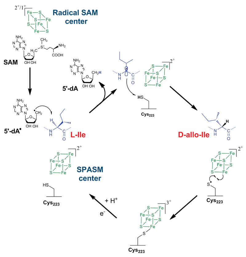
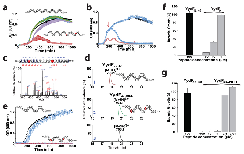
Ribosomally synthesized peptides are built out of L-amino acids, whereas D-amino acids are generally the hallmark of non-ribosomal synthetic processes. Here we show that the model bacterium Bacillus subtilis is able to produce a novel type of ribosomally synthesized and post-translationally modified peptide that contains D-amino acids, and which we propose to call epipeptides. We demonstrate that a two [4Fe-4S]-cluster radical S-adenosyl-L-methionine (SAM) enzyme converts L-amino acids into their D-counterparts by catalysing Cα-hydrogen-atom abstraction and using a critical cysteine residue as the hydrogen-atom donor. Unexpectedly, these D-amino acid residues proved to be essential for the activity of a peptide that induces the expression of LiaRS, a major component of the bacterial cell envelope stress-response system. Present in B. subtilis and in several members of the human microbiome, these epipeptides and radical SAM epimerases broaden the landscape of peptidyl structures accessible to living organisms.


Conflict of interest statement

Competing financial interests

The authors declare no competing financial interests

Figures

Figure 1
Figure 1. YydG is a radical SAM enzyme catalyzing the modification of the YydF peptide.
(a) Structure of the yydFGHIJ operon. yydF: putative peptide, yydG: radical SAM enzyme, yydH: protease, yydIJ ABC-type transporter. (b) Gel electrophoresis analysis of purified YydG expressed in E. coli. (c) UV-visible spectrum of anaerobically reconstituted YydG (blue line) and reduced with sodium dithionite (red line). The symbol * indicates absorbance due to reduced sodium dithionite. (d) Sequence of the YydF peptide from B. subtilis. In grey, region with a low conservation; in blue, strictly conserved amino acid residues (see Supplementary Fig. S2). (e) HPLC analysis of YydF18-49 incubated with YydG after 90 min under anaerobic conditions in the absence (upper trace) or the presence (lower trace) of sodium dithionite as one-electron donor. In the absence of sodium dithionite (upper trace), only the YydF18-49 peptide substrate was monitored. The m/z of each peptide is indicated above the corresponding peaks. See Supplementary Information for experimental conditions. (f) HPLC analysis of SAM incubated with YydG and YydF18-49 after 90 min under anaerobic conditions in the absence (upper trace) or in the presence (lower trace) of sodium dithionite. See Supplementary Information for experimental conditions.
Figure 2
Figure 2. YydG catalyzes H-atom transfer to the peptide backbone.
Tryptic peptide mapping and LC-MS analysis of (a) YydF18-49 or (b) YydF18-49 after incubation with YydG. Numbers indicate the m/z value for each peptide. Sequences in green indicate the relevant peptide identified by LC-MS (i.e. Peptide 1: Ac-GLLDESQK, [M+H]+ = 931.7 ; Peptide 2: VNDLWYFVK [M+2H]2+ =592.6 and Peptide 3: WILGSGH-NH2, [M+H]+ =768.6). Sequences in pink represent the peptides modified by YydG. (c) LC-MS analysis of the peptide YydF18-49 after incubation with YydG in deuterated buffer. (d) Tryptic peptide mapping and LC-MS analysis of YydF18-49 after incubation with YydG in deuterated buffer. (e) LC-MS/MS analysis of Peptide 2 (upper left panel), Peptide 2* (lower left panel), Peptide 3 (upper right panel) and Peptide 3* (lower right panel) obtained after tryptic hydrolysis of YydF18-49 incubated with YydG in deuterated buffer (see full assignment in Supplementary Table S2-5).
Figure 3
Figure 3. YydG catalyzes amino acid epimerization.
LC-MS/MS analysis of (a) L-/D-Leu and L-Ile/D-allo-Ile or (b) L-/D-Val. Upper traces were obtained with standard amino acids while lower traces correspond to amino acids obtained after incubation of YydF18-49 with the radical SAM enzyme YydG in deuterated buffer. Amino acids were analyzed after hydrolysis and derivatization by N-α-(2,4-dinitro-5-fluorophenyl)-L-valinamide (L-FDVA). MS spectra of (c) L-Ile-FDVA, (d) L-Val-FDVA (upper traces) and their D-epimers (lower traces) obtained after incubation of YydF18-49 with YydG in deuterated buffer. Mass spectra correspond to the dominant ions fragments: m/z= 366 and 352 for Ile- and Val-FDVA derivatives, respectively. The amino acids were derivatized by L-FDVA and detected by LC-MS after ion current extraction in MS/MS experiments using the transition 412>366 and 398>352 for Ile/Leu and Val-FDVA derivatives, respectively. (e) LC-MS/MS analysis of the deuterated peptide YydF18-49-VD8 incubated with YydG. After incubation with YydG, YydF18-49-VD8 ([M+2H]2+ = 1887.7) (trace 1) was converted in peptides containing one D-valine ([M+2H]2+ = 1887.2) (traces 2&3) or two D-Val [M+2H]2+ = 1886.7 (trace 4). In the sequences, the octadeuterated L-valine residues are labeled in light blue and the heptadeuterated D-valine in red. Masses indicate mono-isotopic ions. (f) LC-MS/MS analysis of 5'-dA produced by YydG incubated with YydF18-49 (upper trace) or YydF18-49-VD8 (lower trace). (g) Time-course production of 5'-dA (blue symbol) and epimerized peptide (red symbol). See supplementary Information for experimental conditions.
Figure 4
Figure 4. Activity of YydG mutants.
(a) Molecular phylogenetic analysis of radical SAM epimerases and representative members of the radical SAM enzyme superfamily: The evolutionary history was inferred by using the Maximum Likelihood method based on the JTT matrix-based mode. The percentage of replicate trees in which the associated taxa clustered together in the bootstrap test (1000 replicates) are shown next to the branches. Initial tree(s) for the heuristic search were obtained by applying Neighbor-Join and BioNJ algorithms to a matrix of pairwise distances estimated using a JTT model, and then selecting the topology with superior log likelihood value (see Supplementary Information for full methodology. (b) Gel electrophoresis analysis of the purified YydG mutants. Mutants AxxxAxxA (A3), C22A, C222A and C223A. (c) UV-visible spectra of A3 (blue trace), C22A (red trace), C222A (green trace) and C223A (purple trace) mutants after anaerobic reconstitution. (d) HPLC analysis of the reaction after incubation of YydF18-49 and each mutant protein. (e) LC-MS analysis of the reaction catalyzed by the wild-type enzyme or the C223A mutant in the absence of DTT. YydG produced peptides at [M+3H]3+= 1258.3 while the C223A mutants produced additional peptides at [M+Na]+ = 603.2861, [M+2H]2+= 1646.3838 and [M+2H]2+= 1125.7 identified as I44Δ1-H49, G18-I44Δ30 and G18-V36Δ30, respectively (see Supplementary Fig. S22-25 and Supplementary Table S7-12 for full analysis and assignment). (f) Radical fragmentation mechanism of the YydF18-49 peptide leading to the production of the I44Δ1-H49 and the G18-I44Δ30. The theoretical masses for the hydrolytic products (I44-H49 & G18-I44) and the experimental masses measured (I44Δ1-H49 & G18-I44Δ30) are indicated (See Supplementary Table S7 for comparison with theoretical products).
Figure 5
Figure 5. Proposed mechanism of the radical SAM peptide epimerase YydG.
YydG generates a 5′-dA• radical which abstracts an amino acid Cα H-atom. A carbon-centred radical is generated and quenched by the thiolate H-atom of Cys223 leading to the formation of a D-amino acid residue. The SPASM [4Fe-4S] center likely assists the radical quenching by reducing the thiyl radical. Further reduction of Cys223 would be required to allow another catalytic cycle.
Figure 6
Figure 6. Activity of the epipeptides on Bacillus subtilis.
(a) Growth of B. subtilis in liquid LB medium in the presence of YydF18-49 or peptides containing one or two epimerized residues in position 36 and 44. B. subtilis was grown in LB medium alone (black squares), in the presence of YydF18-49 (white squares), YydFa (purple squares), YydFc (Green squares) or YydFb (red squares). Each measurement is the mean of three growth experiments with the SD indicated. (b) Growth of B. subtilis in the presence of YydF18-49 at initial time or mid-exponential phase. B. subtilis was grown in LB medium alone (black squares), in the presence of YydF18-49 (white squares), YydFb (red squares) or after addition of YydFb at mid-exponential phase (indicated by the red arrow) (light blue squares). Each measurement is the mean of three growth experiments with the SD indicated. (c) LC-MS/MS analysis of the purified peptide from B. subtilis. (see Supplementary methods for peptide purification). (d) LC-MS analysis of YydF33-49, YydF33-49DD synthetic peptides (traces 1&2) and the peptide isolated from B. subtilis (trace 3). (See Supplementary Fig. S28 & S29 for complete assignment). (e) Growth of B. subtilis in liquid LB medium in the presence of YydF33-49DD. B. subtilis was grown in LB medium alone (black squares), in the presence of YydF33-49 (white squares) or YydF33-49DD (red squares). Each measurement is the mean of three growth experiments with the SD indicated. Growth ratio of B. subtilis in the presence of (f) YydF18-49 (100 µM) or YydFb and (g) YydF33-49 or YydF33-49DD. Ratios were determined by comparison with growth in the absence of peptide.

References

    1. Jordan S, Junker A, Helmann JD, Mascher T. Regulation of LiaRS-dependent gene expression in bacillus subtilis: identification of inhibitor proteins, regulator binding sites, and target genes of a conserved cell envelope stress-sensing two-component system. J Bacteriol. 2006;188:5153–66. - PMC - PubMed
    1. Jordan S, Hutchings MI, Mascher T. Cell envelope stress response in Gram-positive bacteria. FEMS Microbiol Rev. 2008;32:107–46. - PubMed
    1. Mascher T, Margulis NG, Wang T, Ye RW, Helmann JD. Cell wall stress responses in Bacillus subtilis: the regulatory network of the bacitracin stimulon. Mol Microbiol. 2003;50:1591–604. - PubMed
    1. Mascher T, Zimmer SL, Smith TA, Helmann JD. Antibiotic-inducible promoter regulated by the cell envelope stress-sensing two-component system LiaRS of Bacillus subtilis. Antimicrob Agents Chemother. 2004;48:2888–96. - PMC - PubMed
    1. Butcher BG, Lin YP, Helmann JD. The yydFGHIJ operon of Bacillus subtilis encodes a peptide that induces the LiaRS two-component system. J Bacteriol. 2007;189:8616–25. - PMC - PubMed

Publication types

MeSH terms