. 2018 Nov 2;9(6):472-479.
doi: 10.1080/21541248.2017.1321614. Epub 2017 Jun 23.

An unstructured loop that is critical for interactions of the stalk domain of Drp1 with saturated phosphatidic acid

Affiliations

An unstructured loop that is critical for interactions of the stalk domain of Drp1 with saturated phosphatidic acid

Yoshihiro Adachi et al. Small GTPases. .

Abstract

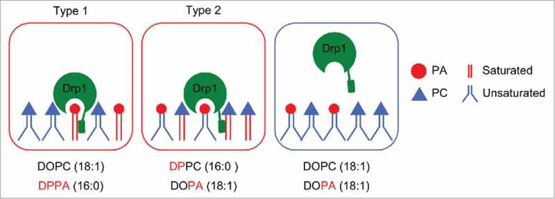
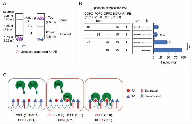
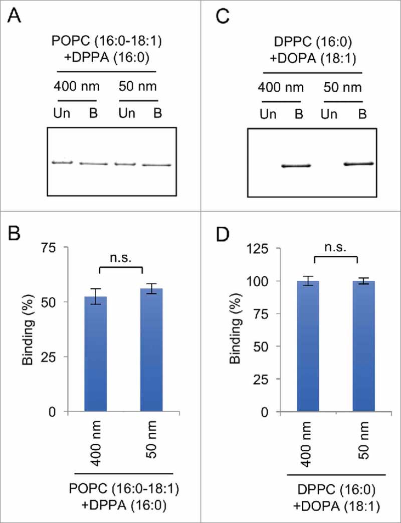
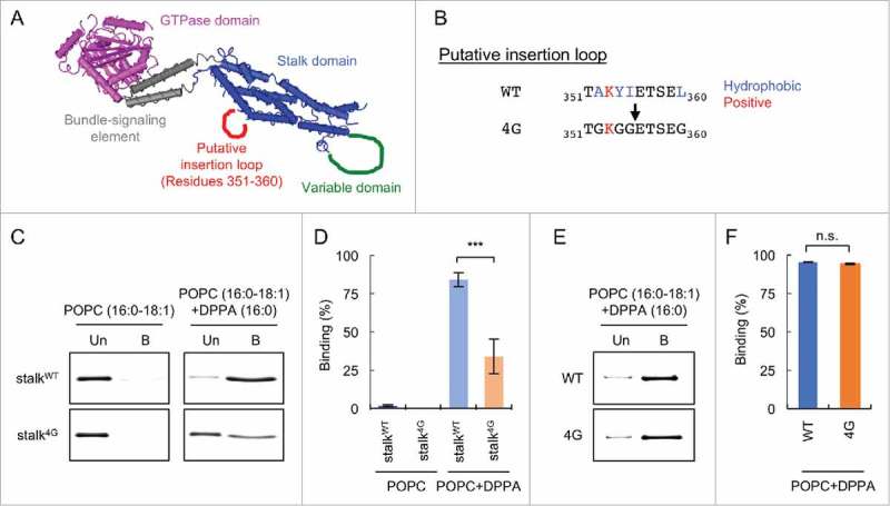
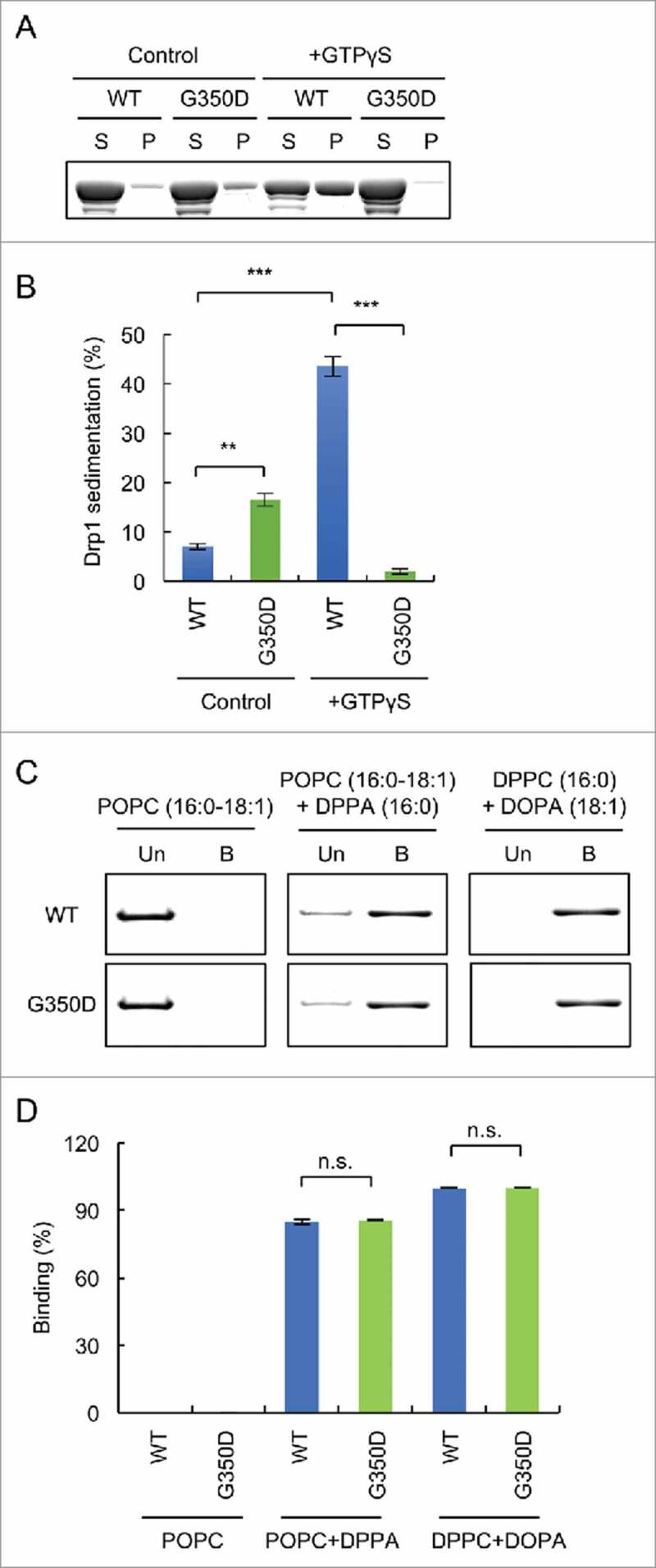
Dynamin-related protein 1 (Drp1) is a dynamin superfamily GTPase, which drives membrane constriction during mitochondrial division. To mediate mitochondrial division, Drp1 is recruited to the mitochondrial outer membrane and is assembled into the division machinery. We previously showed that Drp1 interacts with phosphatidic acid (PA) and saturated phospholipids in the mitochondrial membrane, and this interaction restrains Drp1 in initiating the constriction of mitochondria. Here, we show that the role of saturated acyl chains of phospholipids is independent of their contribution to the membrane curvature or lipid packing suggesting their direct interaction with Drp1. We further show that an unstructured loop in the stalk domain of Drp1 is critical for interaction with unsaturated PA. Our data significantly advance our understanding of this unique protein-lipid interaction involved in mitochondrial division.

Keywords: GTPase; dynamin-related protein 1; lipid binding; liposomes; mitochondria; organelle dynamics; phosphatidic acid.


Figures

Figure 1.
Figure 1.
Coincident interaction of Drp1 with PA. In type 1 binding mode, Drp1 binds to saturated PA. In type 2 binding mode, Drp1 simultaneously binds to unsaturated PA and saturated non-PA phospholipids such as PC.
Figure 2.
Figure 2.
Two saturated acyl chains in single phospholipids are necessary for interactions of Drp1 with liposomes. (A) Liposome flotation assay. Recombinant His6-Drp1 was incubated with liposomes at 4°C for 1 hour and analyzed by sucrose density gradient. The majority of liposomes floated to the upper half of the gradient. (B) Liposome flotation assay was performed using full length His6-Drp1 and the indicated liposomes. Equal amounts of the bottom (unbound, Un) and top (bound, B) were analyzed by SDS-PAGE and silver staining. Band intensity was quantified, and relative amounts of Drp1 in the bound fraction are shown (Mean ± SEM; n = 3). (C) Two saturated acyl chains must be present in the same molecule to mediate Drp1 interactions. Drp1 does not bind to PA that contains one saturated acyl chain and one unsaturated acyl chain. Student's t-test: ***p < 0.001.
Figure 3.
Figure 3.
Drp1 binds to liposomes regardless of their diameter in liposome flotation assays. (A and C) Flotation assays were performed using His6-Drp1 and liposomes that contain saturated PA (DPPA) in (A) and unsaturated PA (DOPA) and saturated PC (DPPC) in (C). To change the diameter, we used 2 different nanopore membranes with a pore size of 50 or 400 nm. The upper and lower fractions were collected and analyzed by SDS-PAGE and silver staining. (B and D) The band intensity was quantified, and the relative amounts of Drp1 in the bound fraction are shown (Mean ± SEM; n = 3).
Figure 4.
Figure 4.
Identification of a loop that is critical for interactions with saturated PA. (A) The 3D structure of Drp136. A putative membrane insertion loop is shown (red). (B) Amino acid sequence of the putative insertion loop. Four hydrophobic residues (blue) are substituted by glycine in the 4G mutant. (C) Liposome flotation assay using His6-stalk and His6-stalk4G. Liposomes containing saturated PA (DPPA) were used. As a negative control, liposomes that lack DPPA were used. (D) Band intensity was quantified, and relative amounts of Drp1 in the bound fraction are shown (Mean ± SEM; n = 3). (E) Liposome flotation assay using His6-Drp1 and His6-Drp14G. (D) Relative amounts of Drp1 in the bound fraction are shown (Mean ± SEM; n = 3). Student's t-test: ***p < 0.001.
Figure 5.
Figure 5.
Drp1 binds to PA-containing liposomes independently of oligomerization. (A) His6-Drp1 and His6-Drp1G350D were incubated in the presence or absence of 2 mM GTPγS at room temperature for 30 min and ultracentrifuged at 100,000 x g for 20 min. The supernatant and pellet fractions were analyzed by SDS-PAGE and Coomassie brilliant blue staining. (B) The band intensity was quantified and relative amounts of Drp1 in the pellet fractions are shown (mean ± SEM; n = 3). (C) Liposome flotation assay using His6-Drp1 and His6-Drp1G350D. (D) Band intensity was quantified, and relative amounts of Drp1 in the bound fraction is shown (mean ± SEM; n = 3). Student's t-test: **p < 0.005; ***p < 0.001.

References

    1. Youle RJ, van der Bliek AM. Mitochondrial fission, fusion, and stress. Science 2012; 337:1062-5; PMID:22936770; https://doi.org/10.1126/science.1219855 - DOI - PMC - PubMed
    1. Pernas L, Scorrano L. Mito-Morphosis: Mitochondrial Fusion, Fission, and Cristae Remodeling as Key Mediators of Cellular Function. Annu Rev Physiol 2016; 78:505-31; PMID:26667075; https://doi.org/10.1146/annurev-physiol-021115-105011 - DOI - PubMed
    1. Friedman JR, Nunnari J. Mitochondrial form and function. Nature 2014; 505:335-43; PMID:24429632; https://doi.org/10.1038/nature12985 - DOI - PMC - PubMed
    1. Shutt TE, McBride HM. Staying cool in difficult times: Mitochondrial dynamics, quality control and the stress response. Biochim Et Biophys Acta 2012; PMID:22683990; https://doi.org/10.1016/j.bbamcr.2012.05.024 - DOI - PubMed
    1. Roy M, Reddy PH, Iijima M, Sesaki H. Mitochondrial division and fusion in metabolism. Curr Opin Cell Biol 2015; 33C:111-8; https://doi.org/10.1016/j.ceb.2015.02.001 - DOI - PMC - PubMed

Publication types

LinkOut - more resources