Review
. 2017 Sep 29;4(1):141-158.
doi: 10.1146/annurev-virology-101416-042001. Epub 2017 Jun 23.

New World Arenavirus Biology

Affiliations
Review

New World Arenavirus Biology

Nicolás Sarute et al. Annu Rev Virol. .

Abstract

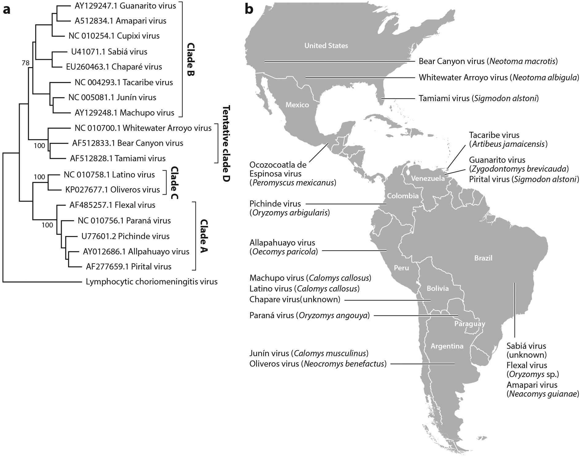
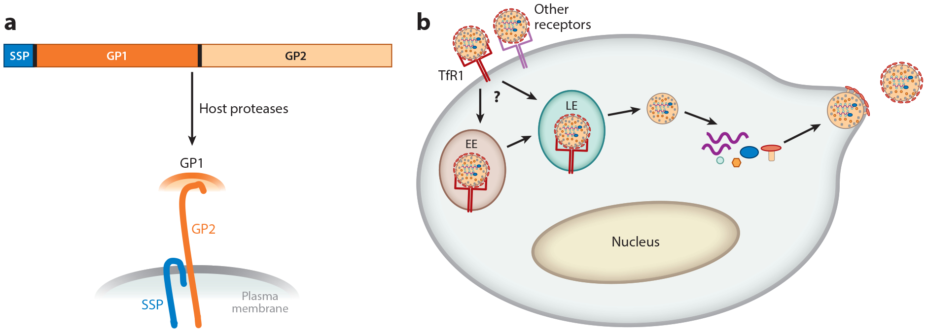
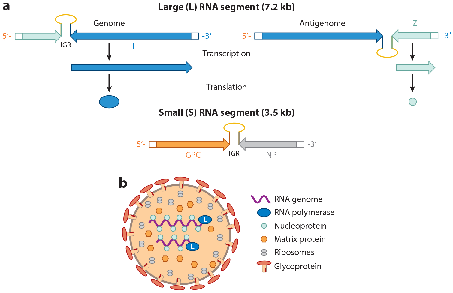
Hemorrhagic fevers caused by viruses were identified in the late 1950s in South America. These viruses have existed in their hosts, the New World rodents, for millions of years. Their emergence as infectious agents in humans coincided with changes in the environment and farming practices that caused explosions in their host rodent populations. Zoonosis into humans likely occurs because the pathogenic New World arenaviruses use human transferrin receptor 1 to enter cells. The mortality rate after infection with these viruses is high, but the mechanism by which disease is induced is still not clear. Possibilities include direct effects of cellular infection or the induction of high levels of cytokines by infected sentinel cells of the immune system, leading to endothelia and thrombocyte dysfunction and neurological disease. Here we provide a review of the ecology and molecular and cellular biology of New World arenaviruses, as well as a discussion of the current animal models of infection. The development of animal models, coupled with an improved understanding of the infection pathway and host response, should lead to the discovery of new drugs for treating infections.

Keywords: animal models; arenavirus; transferrin receptor.


Figures

Figure 1
Figure 1
New World arenavirus taxonomy and geographic distribution. (a) Cladogram inferred by using the maximum-likelihood method with the Kimura two-parameter and gamma distribution substitution model. Phylogenetic reconstruction was performed using the full-length glycoprotein precursor (GPC) gene. Bootstrap values are shown for the main nodes. The GenBank accession number and origin of each sequence are denoted, as are the clade names. A strain of lymphocytic choriomeningitis virus was used as outgroup. (b) Map of the Americas showing the distribution of the New World arenaviruses. Virus names and most frequent host reservoirs are indicated in the boxes.
Figure 2
Figure 2
Cellular infection pathway and structure of the arenavirus glycoprotein (GP). (a) The arenavirus glycoprotein precursor (GPC) is translated as a polyprotein, which is synthesized in the endoplasmic reticulum, modified in the Golgi, and then cleaved by the SK1/S1P cellular protease and host signal peptidase to generate the three subunits—SSP, GP1, and GP2—found in the virion membrane. (b) The pathogenic New World arenaviruses enter cells after binding to transferrin receptor 1 (TfR1) and perhaps other cell surface molecules. After endocytosis to either the early (EE) or late (LE) acidic endosome, which may be cell type dependent, the viral and host membranes fuse and the capsid is released into the cytoplasm. Transcription of the viral RNA follows, to generate the viral genome as well as mRNAs that encode the viral proteins. After synthesis of the proteins, the viral genomic RNAs are packaged into capsids, and the new viruses bud from the plasma membrane, which already contains the viral GP components.
Figure 3
Figure 3
Arenavirus genome and virion structure. (a) Genome organization of the two RNA subunits packaged into arenavirus particles. The large (L) RNA encodes the matrix (Z) and polymerase (L), and the small (S) RNA encodes the glycoprotein precursor (GPC) and nucleoprotein (NP). Both RNAs are ambisense. The scheme for generating the mRNAs for the L and Z RNAs is shown; a similar process occurs for the GPC and NP RNAs. The intergenic region (IGR) between the RNAs controls both RNA transcription and translation. (b) Diagram of the assembled viral particle.

References

    1. Stenglein MD, Sanders C, Kistler AL, Ruby JG, Franco JY, et al. 2012. Identification, characterization, and in vitro culture of highly divergent arenaviruses from boa constrictors and annulated tree boas: candidate etiological agents for snake inclusion body disease. mBio 3:e00180–12 - PMC - PubMed
    1. Radoshitzky SR, Bao Y, Buchmeier MJ, Charrel RN, Clawson AN, et al. 2015. Past, present, and future of arenavirus taxonomy. Arch. Virol 160:1851–74 - PubMed
    1. Peralta LA, Laguens RP, Cossio PM, Sabattini MS, Maiztegui JI, Arana RM. 1979. Presence of viral particles in the salivary gland of Calomys musculinus infected with Junin virus by a natural route. Intervirology 11:111–16 - PubMed
    1. Cajimat MN, Milazzo ML, Bradley RD, Fulhorst CF. 2012. Ocozocoautla de Espinosa virus and hemorrhagic fever, Mexico. Emerg. Infect. Dis 18:401–5 - PMC - PubMed
    1. Abraham J, Kwong JA, Albarino CG, Lu JG, Radoshitzky SR, et al. 2009. Host-species transferrin receptor 1 orthologs are cellular receptors for nonpathogenic new world clade B arenaviruses. PLOS Pathog. 5:e1000358. - PMC - PubMed

Publication types

MeSH terms